

 Sample Submission Guidelines

Matched Tissue–Organoid–Normal Sequencing: Experimental Design and Interpretation
Meta Intent: A practical research guide for designing matched tissue–organoid–normal sequencing studies that distinguish shared tumor biology, sampling heterogeneity, culture selection, and analytical artifacts before a model is used for downstream discovery.
Patient-derived organoids can provide a renewable experimental system, but an organoid is not automatically a molecular duplicate of the specimen from which it was derived. A comparison between one tissue sample and one organoid culture can be useful; it is also easy to over-interpret. A variant can appear only in the organoid because a low-frequency tissue clone expanded in culture. It can appear only in tissue because the source specimen contained subclones, stromal DNA, or normal epithelium that the culture did not retain. A call can move in either direction because of purity, copy number, coverage, or filtering choices. The practical question is therefore not simply, "Do the tissue and organoid match?" It is, "What does each discordance mean, and is the evidence adequate for the specific downstream research decision under review?"
This article sets out a research-use-only framework for answering that question through a deliberately matched triad: source tissue, derived organoid, and a defined normal comparator. It focuses on experimental design and interpretation rather than on assigning a single fidelity score. Before sequencing, basic model identity and contamination checks should already be in place through a sequencing-based organoid qualification framework. The triad then adds the evidence required to interpret somatic variants and molecular shifts with context.
Research boundary: For research use only. This framework supports research model qualification and experimental interpretation, not diagnostic procedures, clinical decision-making, patient stratification, therapeutic selection, or clinical trials.
Why Pairwise Tissue–Organoid Comparison Is Not Enough
A tissue-versus-organoid analysis compares two mixtures produced by very different processes. Tissue DNA may contain malignant cells, normal epithelium, immune cells, fibroblasts, necrosis, and variable amounts of infiltrating blood. Organoid DNA is usually more epithelial, but it may represent a selected subset of the source lesion. Without a matched normal, a variant caller must infer whether observed changes are somatic, germline, recurrent technical artifacts, or population polymorphisms. That uncertainty changes both the call set and the story attached to it.
The normal comparator is not an administrative extra. It is the reference that anchors somatic interpretation, helps detect sample swaps, and makes it possible to review a shared site even when one sample falls below a hard calling threshold. A tissue–organoid–normal design therefore supports three related comparisons: tumor tissue versus normal for the source landscape, organoid versus normal for the cultured landscape, and tissue versus organoid for preservation, selection, or divergence.
Reported concordance is highly sensitive to its denominator. "Shared variants divided by tissue calls," "shared variants divided by the union of calls," "shared driver events," and "correlation of variant allele frequencies" are not interchangeable results. State the denominator, genomic territory, caller filters, and whether the comparison was made at independently called sites or after union-site review. A model can preserve key trunk events while missing low-frequency branch events; it can also show strong global agreement but differ in a pathway that matters to the stated experiment.
 Figure 1: A matched tissue–organoid–normal triad separates source-tissue composition, culture selection, and germline background before variant interpretation.
Define the Matched Biological Unit Before Collection
"Matched" should describe a documented biological relationship, not only a shared patient identifier. Before collection, define the lesion, anatomical region, sampling time, treatment status, and whether the tissue fragment used for organoid establishment is adjacent to the fragment allocated for DNA and RNA. A broad lesion label can conceal regional heterogeneity. If the project asks whether an organoid preserves a dominant clone, one well-defined source region may be appropriate. If the project asks whether the culture captures spatial diversity, collect and label multiple regions rather than averaging them later.
Build a specimen map that follows every aliquot: acquisition time, transport condition, gross orientation, histology block, organoid derivation fraction, nucleic-acid extraction fraction, and freeze-thaw history. This map often resolves apparent biological disagreement. For example, an organoid derived from a viable epithelial-rich edge should not be interpreted as a direct molecular replica of a bulk DNA aliquot enriched for central necrosis and stromal material.
Use histology as an interpretive companion, not merely an intake record. A matched organoid histology workflow can document epithelial architecture, tumor content, and relevant source-to-model differences at the point where samples are allocated. For studies beginning from a new tissue source, the practical milestones in organoid model development should be linked to the baseline specimen ID and passage number that will later appear in sequencing metadata.
Choose the Matched Normal Comparator Deliberately
Peripheral blood is often a useful germline comparator because it is physically separate from the lesion and typically yields ample DNA. It is not automatically perfect: hematologic involvement, clonal hematopoiesis, transfusion history, or low DNA quality can require additional review. Adjacent non-malignant tissue can be informative for tissue context, but it may contain occult tumor cells or field changes. Buccal or saliva specimens can introduce other cell mixtures. A normal organoid may be valuable as a tissue-matched biological reference, but it should not be treated as an unquestioned substitute for constitutional DNA without project-specific evidence.
Record why the comparator was selected, what it is expected to control, and which limitations remain. If adjacent normal is used, preserve pathology review and a contamination screen as explicit deliverables. If blood is used for germline subtraction while adjacent tissue is retained for a microenvironment question, label those functions separately. This discipline prevents a later analysis from silently repurposing a comparator beyond what it can support.
Build a Specimen and Aliquot Map Before Sequencing
Triad studies fail most often at handoffs, not at variant annotation. A practical minimum is one accession key that connects the tissue block, organoid culture, passage, extraction, library, lane, and analysis batch. The same record should note whether the tissue and organoid were sequenced from fresh material, frozen material, or a later culture expansion. When RNA is included, document the extraction chemistry and input integrity for all members of the triad. This is especially important because tissue RNA reflects a multicellular microenvironment while organoid RNA commonly reflects the epithelial compartment.
Pre-allocate enough source material for one confirmatory analysis. It is inefficient to discover an important tissue-only or organoid-only event after all material has been consumed. A small residual DNA aliquot, retained frozen tissue, or a validated targeted panel can provide an escalation path. For studies requiring cross-modal interpretation, organoid sequencing and analysis can be planned around shared sample identifiers rather than as separate DNA and RNA workstreams.
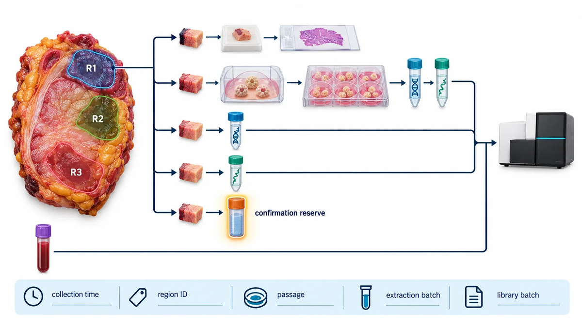 Figure 2: A pre-sequencing aliquot map preserves the link between tissue region, pathology review, organoid passage, normal comparator, library batch, and confirmation reserve.
Select the Sequencing Strategy Around the Interpretation Goal
Start from the result that must be defended. A targeted panel is appropriate when the project asks whether a defined set of established events is retained or when a small number of discordant sites need confirmation. Whole-exome sequencing is useful for coding-region somatic burden, driver analysis, and broader mutation concordance. Whole-genome sequencing is most informative when structural variants, noncoding events, global copy-number architecture, or complex rearrangements matter to the biological question. The relevant goal is not maximum data volume; it is whether the selected assay can discriminate the alternatives in the study hypothesis.
For coding-region questions, an exome sequencing strategy should be designed with equivalent capture chemistry and callable-region criteria across tissue, organoid, and normal. For a question that depends on complex rearrangements or genome-wide copy-number patterns, whole-genome sequencing may provide a more coherent evidence layer. If the priority is a small set of high-consequence sites, a targeted region sequencing approach can be more efficient for replicate or follow-up assessment. These choices should be made before a concordance percentage is promised.
RNA-seq adds a different question: whether the cultured epithelial program and the source tissue program are comparable after accounting for cellular composition. It should not be used as a surrogate for DNA-based somatic calling. If the project needs both molecular layers, keep the DNA and RNA comparison plans distinct and then reconcile their results through a pre-specified interpretation table.
Harmonize Wet-Lab and Bioinformatics Variables
Sequencing triads with different library preparation, capture systems, read lengths, reference genomes, or caller versions creates avoidable discordance. Harmonize what can be harmonized: extraction inputs, library class, sequencing mode, reference build, interval files, alignment method, duplicate handling, and somatic caller settings. When a difference cannot be avoided, record it and restrict direct comparison to regions that are callable in all three samples.
Run identity checks before interpreting biology. Germline fingerprinting markers, sex-chromosome consistency, and pairwise relatedness should be reviewed alongside sample-sheet metadata. Then use a common analysis workflow for tissue-normal and organoid-normal comparisons, followed by union-site review. A variant calling workflow is more defensible when its filters are transparent and the same evidence fields are retained for every member of the triad: depth, alternate reads, base quality, mapping quality, strand representation, local sequence context, and filter status.
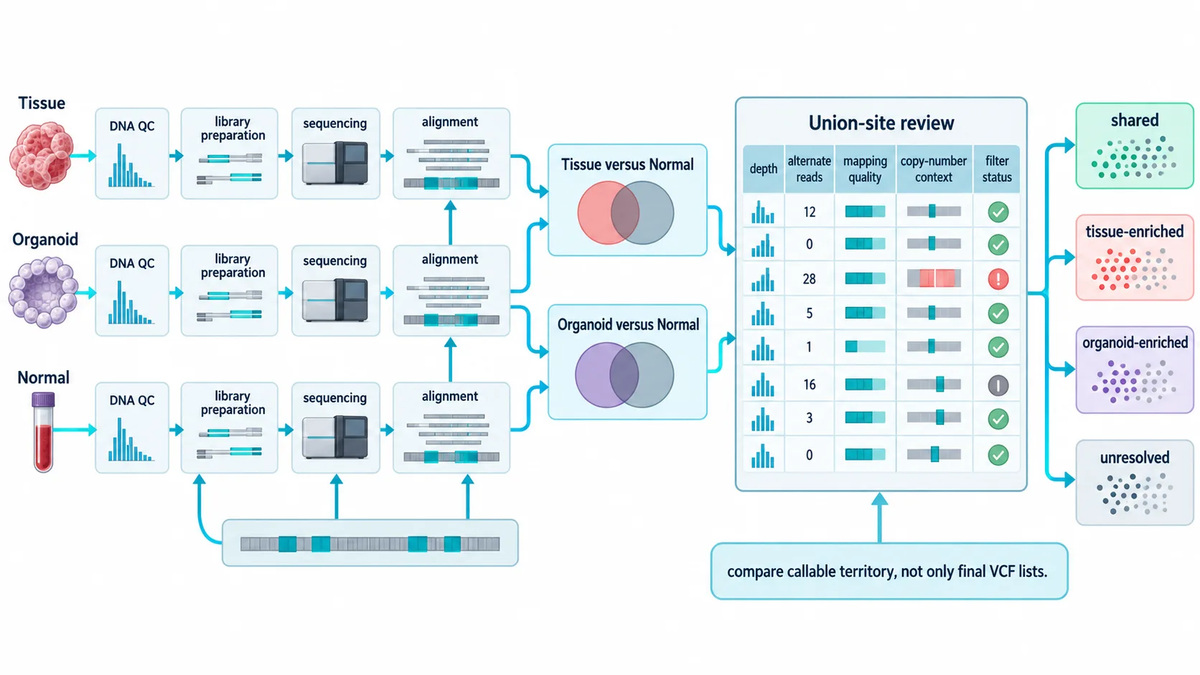 Figure 3: A harmonized analysis path uses matched preprocessing and dual paired calls before union-site review of tissue-only, organoid-only, and shared events.
Interpret Variants with a Three-Way Evidence Grid
Classify every high-priority event by evidence across all three samples rather than by presence or absence in two final VCF files. The key categories are shared somatic events, tissue-enriched events, organoid-enriched events, germline or likely germline events, and unresolved events. The unresolved category is important. A site below a calling threshold in one sample may still show supporting reads, or it may lie in a poorly callable region. Neither situation supports a clean biological conclusion.
| Observed pattern | Common explanation | Next interpretive step |
|---|---|---|
| Present in tissue and organoid; absent from normal | Shared somatic trunk event | Review depth and local quality; assess copy-number context before comparing VAF. |
| Strong in tissue; weak or absent in organoid | Spatial subclone, low organoid representation, or tissue-only component | Inspect organoid raw evidence and source-region histology; confirm if the event changes model suitability. |
| Weak in tissue; strong in organoid | Culture enrichment of a pre-existing clone, tissue purity difference, or calling asymmetry | Perform union-site review; consider targeted confirmation of the source tissue. |
| Present in all three samples | Germline event, mapping issue, or normal contamination context | Review population frequency, normal depth, local alignment, and comparator suitability. |
The aim is not to force every variant into a tidy category. It is to ensure that the evidence behind a model decision is visible. A curated shared-driver list, a copy-number comparison, and a documented set of consequential discordances are usually more useful than an unqualified percentage of "matching variants."
Do Not Compare Raw VAF Without Purity and Copy-Number Context
Variant allele fraction is a measurement, not a direct measure of clonality. Tissue VAF is shaped by tumor purity, local copy number, mutation multiplicity, and subclonal composition. An organoid may display a higher VAF simply because non-epithelial cells are depleted in culture, not because the mutation arose or became biologically dominant. Conversely, a lower organoid VAF can reflect copy-number change, sampling, or a mixed culture.
Review VAF alongside purity estimates and copy-number profiles. A copy-number sequencing analysis can reveal whether an apparent VAF shift is compatible with a gain, loss, or allelic imbalance at the locus. When purity estimates are uncertain, report VAF patterns descriptively and avoid converting them into definitive clone frequencies. This article complements the matrix guide on detecting normal-cell overgrowth before sequencing: that guide asks whether model composition is a pre-sequencing risk, while the present triad asks how composition changes the meaning of observed variants.
A useful review order is to first ask whether the locus is callable in every sample, then whether the observed VAF pattern is plausible under the local copy-number state, and only then whether a clonal interpretation is warranted. This sequence avoids a common error: treating a visually striking allele-fraction difference as evidence of evolutionary change before technical and compositional explanations have been considered. If no reliable purity or copy-number estimate is available, the conclusion should remain limited to the observed sequencing evidence.
Distinguish Source Heterogeneity from Culture Selection
Not all mismatch is drift. Tissue may contain multiple spatial clones, and the organoid may preferentially establish from one viable epithelial compartment. Culture can also select cells according to medium, matrix, passage, and dissociation stress. To distinguish these mechanisms, review the timing and pattern of change. A variant detectable at low level in the baseline tissue and enriched in early passage supports selection from the source. A new event that is absent after sensitive review of tissue, early passage, and normal deserves a higher level of scrutiny, but still requires technical confirmation before it is described as culture-acquired.
Use longitudinal samples when the question is stability rather than baseline fidelity. The matrix article on passage, matrix, and culture-medium effects on organoid drift provides the experimental controls for assigning that cause. Here, the matched normal is retained so that later comparisons remain somatically anchored. It is particularly valuable when a culture is expanded, thawed, or exposed to an experimental perturbation before downstream sequencing.
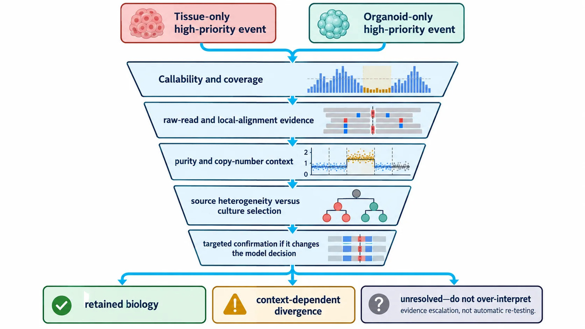 Figure 4: Discordant variants are triaged through callability, raw-read evidence, purity and copy-number context, source heterogeneity, and targeted confirmation before biological interpretation.
Validate Only the Discordances That Can Change the Model Decision
Confirmation is most valuable when a discordance changes whether the model can answer the stated question. Examples include loss of a defining driver event, an organoid-enriched resistance-associated pathway event relevant to the research hypothesis, a copy-number change that changes dosage interpretation, or a suspected sample identity problem. A focused follow-up using targeted region sequencing can test a small number of sites across the retained tissue, organoid, normal, and longitudinal passages. Orthogonal approaches may be useful for selected sequence changes, but should be planned around the type of uncertainty rather than applied automatically.
Do not validate every low-confidence discrepancy. First remove sites explained by low depth, low mapping quality, repetitive sequence, or non-overlapping capture territory. Then rank remaining sites by model-decision impact. This concentrates material and budget on the evidence that will change the research plan.
Minimum Reporting Package for a Matched Triad Study
A reusable report should allow another researcher to understand what was compared and what was not. Include: sample provenance and tissue-region map; normal-comparator rationale; organoid passage and culture context; pathology or morphology observations; library and analysis harmonization details; callable-region criteria; identity-check result; purity and copy-number context; shared and discordant high-priority events; confirmation status; and an explicit conclusion tied to the intended use of the model.
When the study includes broader fidelity questions, direct readers to the matrix framework on whether patient-derived organoids still match the original tissue. That framework compares multiple molecular layers. The matched-triad report should remain narrower: it documents whether the tissue, culture, and comparator were designed and analyzed well enough to support somatic interpretation.
Make Concordance Metrics Answer a Specific Question
A single concordance value can hide more than it reveals. Before calculating one, decide what it is intended to describe. For source representation, report the proportion of source-tissue somatic events recovered in the organoid within jointly callable regions. For overall overlap, report the shared proportion of the union set. For model suitability, report retention of the pre-specified driver, copy-number, structural, or pathway events that define the research question. These metrics can coexist, but they should not be collapsed into a single claim of "fidelity."
Separate high-confidence events from exploratory findings. A high-confidence comparison has adequate depth in all three samples, consistent mapping and base-quality evidence, and a clear filter status. An exploratory site may be biologically interesting but lack enough data in one member of the triad. Label it accordingly rather than allowing it to inflate either discordance or concordance. This is particularly important in heterogeneous tissue, where a true low-frequency source clone can sit close to the detection limit.
Use a small visual evidence table for the events that matter most: one row per event, with tissue and organoid allele fractions, local depth, normal evidence, copy-number state, tissue-content estimate, classification, and confirmation status. Include a concise interpretation note, such as "shared trunk event," "tissue-enriched, supported by source heterogeneity," or "organoid-enriched, requires confirmation." This transforms the report from a static comparison into a decision record that can be revisited when the same culture is expanded or used in a different experiment.
Set Escalation Rules Before Seeing the Results
Pre-specified escalation rules reduce confirmation bias. A simple rule set can state that a discordance is reviewed further when it affects a model-defining driver, a planned experimental biomarker, a copy-number event that changes dosage interpretation, or an identity and contamination control. For each trigger, define the preferred evidence upgrade: targeted re-sequencing, a second source-region aliquot, another early-passage DNA extraction, an orthogonal sequence assay, or review of the culture record. The rule should also state when no escalation is needed, such as a low-confidence site outside the decision scope.
Longitudinal sampling deserves the same discipline. Baseline, post-expansion, post-thaw, and post-treatment samples should be compared against a defined baseline triad rather than against whichever prior dataset is most convenient. The timing framework in When Should Organoids Be Sequenced? can be used to select checkpoints. The present framework then determines how any observed change is interpreted against the original tissue and matched normal.
A Practical Matched Tissue–Organoid–Normal Workflow
- Write the decision question. Specify the events, pathways, or model properties that must be preserved for the planned study.
- Define the biological unit. Label lesion, region, collection time, and tissue fractions before organoid derivation.
- Select the normal comparator. State what it controls and any known limitations.
- Reserve material. Preserve DNA or tissue for a high-impact discordance check.
- Choose the assay intentionally. Match targeted, exome, or genome-scale sequencing to the interpretation goal.
- Harmonize processing. Use common references, intervals, pipelines, and callable-site rules wherever possible.
- Review a three-way evidence grid. Evaluate shared, tissue-enriched, organoid-enriched, germline, and unresolved sites.
- Escalate selectively. Confirm only discordances that change the model decision, then document the conclusion and its limits.
Frequently Asked Questions
Is blood always the best matched normal?
No. Blood is often useful for germline comparison, but the appropriate comparator depends on disease context, specimen quality, and the question being asked. Define its intended role before sequencing.
Can an organoid-only mutation be called culture-acquired?
Not from an independent call list alone. Review the site in source tissue, normal, and early-passage material; assess callability and raw evidence; then use targeted confirmation if the result matters to model use.
What concordance metric should be reported?
Report the metric that fits the question, but always state its denominator, genomic territory, filters, and whether union-site review was used. A shared-driver metric and a global somatic concordance metric answer different questions.
Why can VAF be higher in the organoid than in the tissue?
Organoid cultures often contain a higher epithelial fraction than bulk tissue. Local copy-number changes and selection of a source subclone can also alter the observed fraction.
Do RNA-seq and DNA sequencing need to use the same tissue fragment?
They need documented spatial and temporal relationships. If exact co-allocation is impossible, record which region and aliquot generated each dataset and avoid treating expression differences as direct proof of DNA divergence.
When should triad sequencing be repeated?
Repeat or extend it when the organoid undergoes a major culture transition, when a model-defining event becomes uncertain, or when longitudinal stability is necessary for the next research stage.
Reference:
- Tsai S, et al. Development of primary human pancreatic cancer organoids, matched stromal and immune cells and 3D tumor microenvironment models. BMC Cancer. 2018;18:335. DOI: 10.1186/s12885-018-4238-4.
- Li X, et al. Organoid cultures recapitulate esophageal adenocarcinoma heterogeneity providing a model for clonality studies and precision therapeutics. Nature Communications. 2018;9:2983. DOI: 10.1038/s41467-018-05190-9.
- Wei L, et al. Pitfalls of improperly procured adjacent non-neoplastic tissue for somatic mutation analysis using next-generation sequencing. BMC Medical Genomics. 2016;9:64. DOI: 10.1186/s12920-016-0226-1.
- Bergmann EA, et al. Conpair: concordance and contamination estimator for matched tumor-normal pairs. Bioinformatics. 2016;32(20):3196-3198. DOI: 10.1093/bioinformatics/btw389.
- Tbeileh N, et al. Metastatic colorectal adenocarcinoma tumor purity assessment from whole exome sequencing data. PLOS ONE. 2023;18(4):e0271354. DOI: 10.1371/journal.pone.0271354.