

 Sample Submission Guidelines

Do Patient-Derived Organoids Still Match the Original Tissue? A Multi-Omics Fidelity Framework
Meta Intent: A practical multi-omics framework for determining which genomic, transcriptomic, and epigenomic features patient-derived organoids retain from source tissue, which differences reflect expected culture biology, and which discrepancies make the model unsuitable for a defined research question.
Patient-derived organoids (PDOs) are often described as faithful models of the tissue from which they were established. That shorthand is useful, but it is incomplete. A PDO may preserve a key driver mutation while losing a low-frequency subclone. It may retain a tumor lineage program while no longer containing the stromal or immune cells that shaped the original bulk-tissue transcriptome. It may cluster with the donor sample by DNA methylation while showing a culture-adapted proliferative RNA program.
Those outcomes are not automatically failures. They become failures only when they undermine the biological claim a project is trying to make. The right question is therefore not "Is this organoid faithful?" but "Faithful to which source feature, at which molecular layer, and for which research decision?" This article provides a practical answer for teams comparing source tissue and PDOs with DNA sequencing, RNA-seq, and epigenomic data.
Fidelity Is Not a Single Similarity Score
A tissue sample is a mixed ecosystem. In a tumor specimen, malignant epithelial cells coexist with fibroblasts, immune populations, endothelial cells, extracellular matrix, necrotic regions, and spatially separated subclones. A conventional PDO is usually enriched for expandable epithelial populations. That enrichment can make some tumor signals easier to detect, but it also means a PDO is not expected to reproduce every tissue-level readout.
For practical model qualification, separate three questions that are frequently blended together:
- Provenance: Does the culture originate from the designated donor specimen?
- Tumor-cell fidelity: Does it retain the tumor-cell-intrinsic genomic architecture and molecular state needed for the study?
- Tissue representation: Does it retain the cellular composition and context required for a tissue-level question?
Provenance is largely an identity question. Fidelity is multidimensional. Representation is use-case specific. A PDO can be highly suitable for studying tumor-cell drivers and still be a poor stand-alone system for questions that require intact immune, vascular, or fibroblast biology. This distinction is the foundation for a defensible organoid research strategy.
 Figure 1: Four Layers of Organoid Fidelity — A comparative framework distinguishing sample provenance, tumor-cell genome, molecular state, and tissue representation.
Start with a Claim-Matched Comparator
A multi-omics comparison is only interpretable if the comparator is defined before data analysis. "Original tissue" may mean a single biopsy core, a resection block, a macrodissected tumor region, or a mixed specimen with variable tumor content. Each represents a different biological denominator. If the tissue contains abundant non-malignant cells, a low bulk-RNA correlation may mainly describe composition rather than tumor-cell divergence.
Before selecting assays, document the source region, pathology estimate, preservation method, organoid passage at harvest, extraction workflow, and whether matched normal material exists. These fields do not create fidelity, but they prevent later disagreement from being attributed to culture when it originated from sampling. Projects that need standardized derivation and traceable starting conditions can connect this record to organoid model development and organoid histology workstreams.
A useful comparator contract contains one sentence: "This PDO will be evaluated for retention of [defined feature] from [defined tissue region] at [defined culture snapshot] to support [defined research use]." That sentence determines the evidence hierarchy. A study of driver architecture should prioritize somatic variants, copy-number states, and clonal structure. A study of pathway activity should prioritize cell-state programs rather than raw gene-level correlation. A study of inflammatory context should explicitly recognize that an epithelial PDO alone cannot be expected to mirror the whole source tissue. Comparative colon and rectal organoid work further supports treating tissue origin and anatomic context as part of the comparator definition, not as background metadata.[5]
Genomic Fidelity: Compare Architecture Before Counting Variants
Genomic comparison should begin with the events most likely to define the model: established driver alterations, focal gains or losses, structural variants, loss of heterozygosity, broad copy-number patterns, and relevant mutational signatures. A long list of shared single-nucleotide variants is helpful, but it is not the entire argument. In many projects, preservation of the tumor's driver configuration and copy-number architecture is more decision-relevant than an absolute percentage of shared calls.
Organoid exome sequencing is a practical starting point when the question is centered on coding drivers and somatic variation. Where structural events, noncoding variants, or ploidy context are important, broader genome-level data may be more appropriate. The analysis should report at least four groups: variants shared by tissue and PDO, variants detected only in tissue, variants detected only in PDO, and variants that are technically indeterminate because of coverage, tumor content, or assay sensitivity.
Private calls should not be treated as proof that new mutations arose in culture. Tissue-only calls may belong to a subclone not represented in the initiated culture, may fall below detection in the PDO, or may be obscured by tissue purity. PDO-only calls may have been present below the tissue detection limit, become easier to observe after epithelial enrichment, or reflect genuine culture selection. The distinction requires variant depth, allele fraction, local copy number, and the expected detection limit—not a binary overlap score alone. Paired bladder PDO work likewise reported retention of key genetic alterations and multiclonal landscapes while documenting case-specific private events and variation in tumor content.[3]
Variant allele fraction (VAF) shifts deserve a separate view. A higher VAF in the PDO can result from increased tumor purity, enrichment of a pre-existing clone, or copy-number change. It should be interpreted alongside copy-number data and not as a direct measure of clonal growth. For projects needing integrated somatic-call review, organoid sequencing analysis can organize tissue–PDO concordance at the event, locus, and pathway levels.
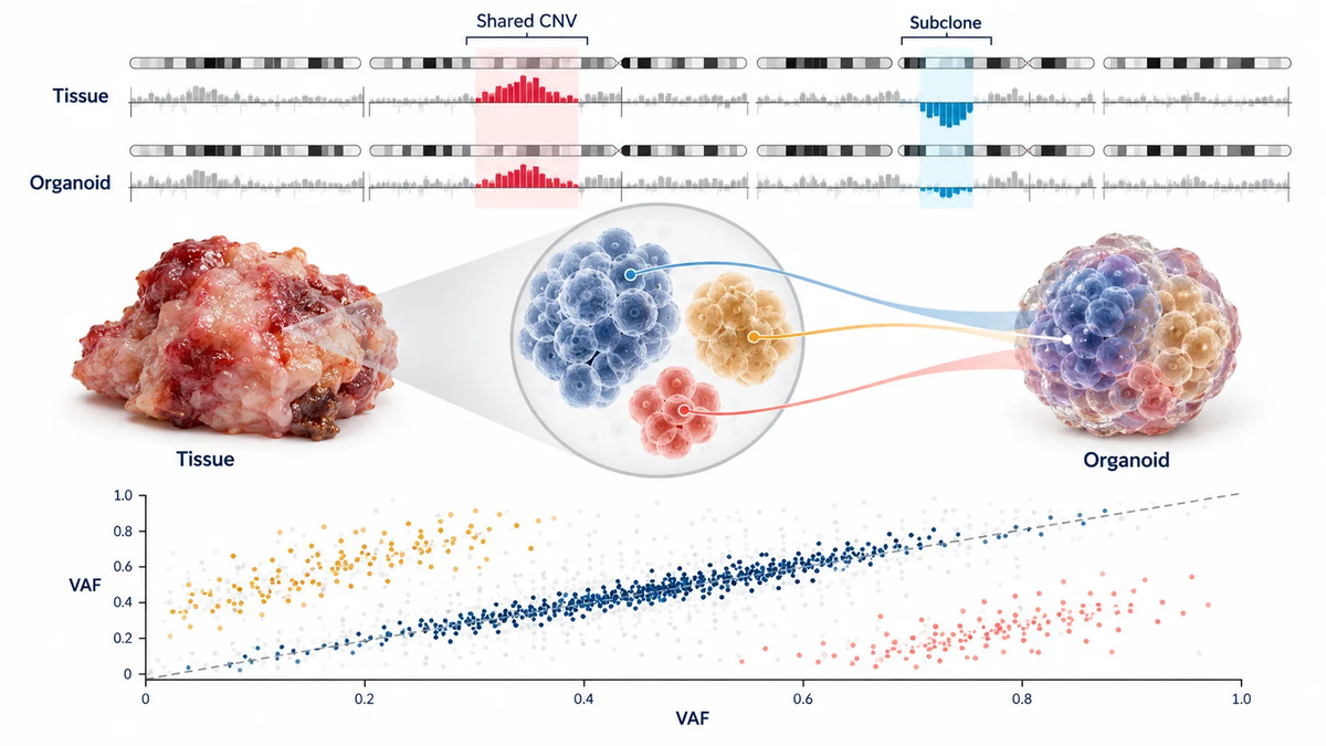 Figure 2: Genomic Concordance and Clone Selection — A matched comparison of shared drivers, copy-number architecture, and variant allele fraction shifts.
Transcriptomic Fidelity: Separate Cell State from Cell Composition
Bulk RNA-seq creates a common interpretation trap. A tissue–PDO pair may show only moderate global correlation because the tissue contains immune and stromal transcripts absent from the culture. Conversely, a high global correlation can hide a meaningful loss of a subtype program if stable housekeeping and lineage genes dominate the statistic. Correlation is a starting point, not the conclusion.
A stronger RNA framework examines three layers. First, ask whether matched samples cluster by donor rather than by sample type. Second, assess whether lineage markers, subtype signatures, and disease-relevant pathways are retained. Third, review the pathways that differ between tissue and PDO and determine whether they are expected composition-dependent programs, culture-associated programs, or changes that challenge the intended model use.
For example, reduced immune signaling, extracellular-matrix transcripts, and vascular signatures in a conventional PDO may be biologically expected. By contrast, loss of the epithelial lineage program, a key pathway activity state, or a molecular subtype signature can be material. The single-cell colorectal study by Wang and colleagues illustrates why this separation matters: tumor-derived organoids retained major tumor-cell programs, while normal-tissue-derived organoids could acquire tumor-like expression features despite retaining normal genomic characteristics.[1]
RNA-seq is sufficient for many state-level questions when paired tissue and organoid samples are processed comparably. When a bulk discrepancy cannot distinguish cell composition from cell-intrinsic state, an orthogonal histology review, deconvolution analysis, or single-cell design may be warranted. That escalation should be driven by the claim—not added by default.
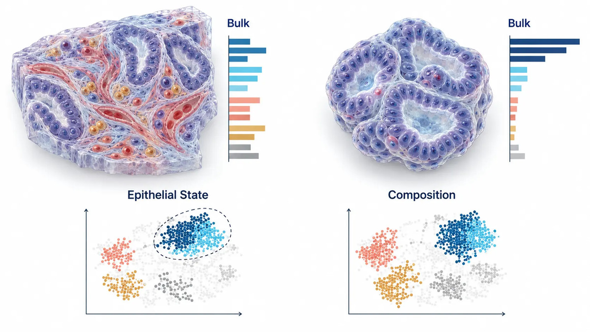 Figure 3: Transcriptomic State versus Cell Composition — Bulk-tissue differences can reflect missing non-epithelial compartments rather than loss of epithelial cell-state fidelity.
Epigenomic Fidelity: Retained Lineage Memory Does Not Mean No Adaptation
DNA methylation and chromatin accessibility can add information when DNA and RNA results do not tell the same story. Epigenomic data may preserve stable lineage or subtype features even when immediate transcriptional responses differ under culture. It can also reveal culture-associated remodeling that is not obvious from driver mutations alone.
The right epigenomic method depends on the uncertainty. Broad DNA methylation profiling is useful for evaluating subtype-associated patterns, patient-specific methylation structure, and candidate differentially methylated regions. Targeted assays are often more efficient when the project concerns defined loci. Chromatin-accessibility approaches are more relevant when the question concerns regulatory-state changes or transcription-factor programs. Organoid epigenetics can be positioned as a focused escalation layer rather than a mandatory assay for every PDO.
Interpret methylation and expression together. A PDO may retain a broad methylation class while showing an RNA shift in proliferation, stress response, or differentiation. That pattern can support lineage retention with culture-dependent state adaptation. In contrast, simultaneous loss of a molecular class, key regulatory features, and corresponding expression programs is more difficult to reconcile as a harmless culture effect. Recent paired model work in glioblastoma illustrates that genomic, epigenomic, and tumor-cell heterogeneity can be retained together, while the abundance of non-malignant components remains variable across models.[2] Paired breast tumor–organoid work similarly shows why DNA methylation and transcriptome comparisons should be interpreted alongside the loss of tumor-microenvironment components.[4] A recent medulloblastoma PDO study likewise illustrates the value of pairing whole-exome, RNA-seq, and methylation evidence when the research question depends on more than one molecular layer.[6]
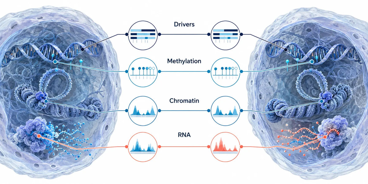 Figure 4: Epigenomic Fidelity and Culture Adaptation — Genomic drivers, methylation state, chromatin accessibility, and RNA programs can retain different levels of concordance.
Read Multi-Omics Discordance as Evidence, Not Noise
The most useful part of a multi-omics comparison is often the disagreement. Averaging all layers into one score can erase the clue that directs the next experiment. Instead, classify the pattern.
Stable genome, shifted transcriptome
This pattern is compatible with altered media signaling, matrix conditions, oxygen exposure, removal of non-malignant cells, or reversible cell-state adaptation. It may still support driver-focused or genetic-perturbation research, but it warrants caution for studies that rely on native tissue-state transcriptional signatures.
Shared drivers, altered VAF structure
This suggests that the core disease genotype is retained but that clone proportions may have changed. The model can be useful for a defined dominant clone, yet it should not be presented as a complete representation of intratumoral diversity. A dedicated organoid sequencing service can help distinguish detection sensitivity from altered clone representation by integrating depth, purity, and copy-number context.
Retained methylation class, altered pathway output
This pattern often points to preserved lineage identity with a shifted functional state. The next action is usually not to reject the PDO. It is to ask whether the shifted pathway is central to the intended assay and whether culture variables, histology, or a targeted perturbation can explain it.
Cross-omics agreement, histology disagreement
If DNA, RNA, and methylation appear consistent but morphology or marker staining conflicts, reassess the tissue region, sectioning plane, sample labeling, and compartment composition. Molecular agreement cannot compensate for a poorly defined tissue comparator.
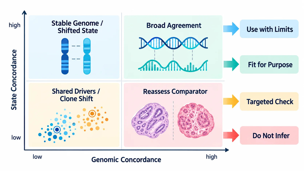 Figure 5: Cross-Omics Discordance Matrix — A decision map for interpreting genomic, transcriptomic, epigenomic, and histologic disagreement between tissue and PDOs.
Build a Purpose-Weighted Fidelity Scorecard
A practical report should not end with "matched" or "unmatched." It should state what evidence is present, what remains uncertain, and whether the model supports the proposed study. A scorecard can include provenance confidence, driver retention, copy-number concordance, clonal representation, lineage and subtype retention, pathway concordance, epigenomic concordance, tissue-composition limitations, and unresolved technical confounders.
These domains should not receive equal weight in every project. A study of a driver alteration may place the greatest weight on somatic calls, CNV, and allele fractions. A study of drug-induced transcriptional adaptation should emphasize baseline and post-perturbation RNA states. A study of epigenetic memory should place methylation or accessibility evidence near the center. A project about immune interactions should explicitly flag the absence of immune composition as a scope limitation rather than silently treating it as a negative data point.
There is no scientifically defensible universal cutoff such as "a correlation above 0.8 proves fidelity." Performance depends on tumor purity, sampling region, assay depth, molecular layer, and the biological claim. Use within-cohort distributions, matched-pair controls where available, and pre-defined decision criteria. This is also where the prior sequencing-based organoid model qualification framework becomes useful: general qualification evidence establishes whether a model can enter research use, while this article determines whether it is representative enough for a specific tissue-linked question.
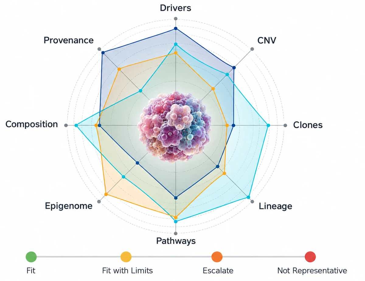 Figure 6: Purpose-Weighted Fidelity Scorecard — Evidence domains are weighted by the defined research use rather than a universal pass threshold.
A Minimum Multi-Omics Workflow for Tissue-to-PDO Comparison
- Define the research claim. State whether the model must retain a driver, a cellular state, an epigenomic class, or a tissue interaction.
- Define the comparator. Record source region, pathology context, tumor content, processing conditions, and PDO harvest snapshot.
- Establish genomic architecture. Compare drivers, CNV, relevant structural events, and allele-fraction patterns.
- Test functional molecular state. Use RNA-seq to assess lineage, subtype, and study-relevant pathways, not only global correlation.
- Add epigenomic evidence when it resolves an actual ambiguity. Use methylation or accessibility data when lineage, regulatory state, or stable memory is central to the claim.
- Classify discrepancies. Decide whether they are expected composition effects, plausible culture adaptation, unresolved technical issues, or disqualifying loss of a required feature.
- Assign a research-use status. Report fit for purpose, fit with stated limitations, requires targeted escalation, or not representative for the intended claim.
This workflow creates a bridge between model generation and data interpretation. It also clarifies when to move into a more targeted investigation, such as tumor-purity assessment before sequencing or a planned matched tissue–organoid–normal sequencing design. Those follow-on questions are related but should not be conflated with the basic fidelity decision.
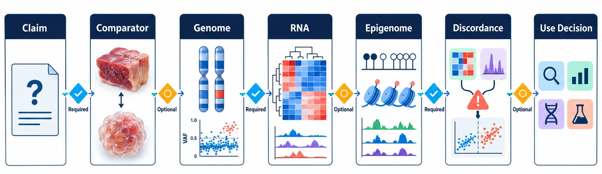 Figure 7: Multi-Omics Fidelity Decision Workflow — A stepwise route from comparator definition through multi-omics comparison, discrepancy classification, and research-use decision.
What Each Evidence Layer Can and Cannot Resolve
Multi-omics does not mean that every assay answers every fidelity question. Treat each layer as a measurement with a defined resolution and a defined blind spot. Histology establishes morphology and the presence or absence of recognizable compartments. DNA sequencing evaluates heritable architecture. RNA-seq reports the molecular state captured at harvest. Methylation and chromatin data can add a more stable regulatory view, but they still depend on sample composition and assay design.
A compact evidence hierarchy helps prevent over-interpretation. Histology can show whether the organoid retains glandular, solid, or mixed morphology, but it cannot establish a complete driver landscape. Exome or genome sequencing can show shared mutations and copy-number patterns, but it cannot tell whether a pathway is transcriptionally active at the time of assay. Bulk RNA-seq can identify altered programs, but it cannot always distinguish state change from the loss of an immune or stromal compartment. Single-cell or spatial analysis can resolve that ambiguity, but it is not automatically necessary for every pair.
The most efficient design is sequential. Start with morphology, DNA evidence, and bulk RNA evidence that directly address the planned use. Escalate only when an unresolved difference would change the interpretation. For example, if a PDO retains a required driver and copy-number pattern but has lower inflammatory RNA signatures than tissue, a full epigenomic panel may not solve the primary uncertainty. A pathology review or an immune co-culture design may be more informative. Conversely, if a key lineage signature is lost in RNA-seq despite retained drivers, a methylation or chromatin assay can test whether the underlying lineage program is preserved but transcriptionally suppressed.
This approach helps teams use multi-omics analysis as a decision tool rather than as a large, undirected panel. It also makes the final report easier to audit: every assay has a stated question, a comparison metric, and a defined action if the result disagrees with the source tissue.
Use the same biological denominator whenever possible
Comparisons are strongest when tissue and PDO data are normalized to a comparable biological compartment. If tissue RNA contains abundant fibroblast, endothelial, and immune populations, compare the PDO against an epithelial-enriched tissue view when possible, or explicitly model the non-epithelial contribution. If a tissue sample has low tumor content, report that as a limitation of somatic-variant and copy-number sensitivity. If the PDO is grown from a small lesion area while the tissue reference came from a distant region of a heterogeneous resection, the result is a regional comparison, not a global tumor comparison.
Do not replace a paired analysis with a general tumor atlas. Reference cohorts are useful for determining whether both tissue and PDO belong to the expected disease class, but they cannot prove pairwise fidelity. A PDO can resemble a cohort subtype and still fail to retain the specific donor feature the project needs. Pairwise evidence should remain primary; external reference data should supply context.
When a Fidelity Result Is Uninterpretable
Some tissue–PDO comparisons should be labeled inconclusive rather than forced into a pass or fail category. This is especially important when analytical limitations could mimic biological divergence. Common examples include low tissue tumor fraction, uneven DNA or RNA quality, non-overlapping tissue regions, an organoid sample harvested after an unrecorded culture change, insufficient sequencing depth at a relevant locus, and a bulk comparison dominated by different cell compositions.
Inconclusive does not mean the model is unusable. It means the available evidence cannot support the exact claim. The appropriate next step is a narrow rescue experiment. For uncertain somatic calls, increase coverage at the critical loci or use a matched-normal-aware analysis. For ambiguous copy-number findings, inspect segmentation quality and allele-specific signals. For transcriptomic discordance, test whether the differences map to non-epithelial genes, stress programs, or the target pathway. For methylation disagreement, distinguish global technical variation from coherent changes at biologically relevant regions.
A strong organoid characterization package should therefore include an uncertainty field, not only a similarity metric. The report should identify the evidence that supports use, the evidence that limits use, and the one most efficient experiment that could resolve the open question. That structure is more actionable than a generic statement that the PDO "closely resembles" the original tissue.
Expected divergence, actionable uncertainty, and disqualifying divergence
Expected divergence is a difference that follows directly from the model format and does not undermine the stated claim. A conventional epithelial PDO will often show lower immune, vascular, stromal, and extracellular-matrix signals than a bulk tissue comparator. It may also show a stronger proliferation program after expansion. These are important scope limitations, but they are not reasons to reject a PDO that is being used to study a retained tumor-cell driver or epithelial response. The report should name the missing or shifted compartment, explain why it is expected, and state which research questions are therefore out of scope.
Actionable uncertainty occurs when the discrepancy might be biological but the available data cannot yet exclude a technical explanation. For example, a tissue-only driver may sit near the assay detection limit; an apparent copy-number difference may reflect low tumor fraction or segmentation quality; and an RNA signature may differ because the tissue contains non-epithelial cells. The appropriate response is a focused follow-up, such as deeper targeted sequencing, pathology-guided region matching, deconvolution, or a matched-normal-aware review. It is usually more informative to resolve the one uncertainty that affects the claim than to add an unfocused large assay panel.
Disqualifying divergence is the loss or replacement of a feature required for the proposed research use. Examples include absence of a required driver or subclone, loss of the molecular subtype that defines the study cohort, a major lineage shift confirmed across orthogonal assays, or failure to retain the epigenomic class that the experiment needs to represent. This classification is not a statement that the PDO has no value. It means that the available model should not be used as evidence for that particular tissue-linked question without a different comparator, additional model, or redesigned study claim.
From Fidelity Evidence to a Defensible Research Statement
The final conclusion should match the evidence layer that was actually tested. If genome-wide copy-number patterns and core drivers are retained, the defensible statement is that the PDO preserves the source tumor's major genomic architecture at the evaluated snapshot. If tumor-cell lineage and pathway signatures are also retained after accounting for composition, the statement can extend to tumor-cell transcriptomic state. If methylation or accessibility data agree, the model has stronger support for retaining a regulatory identity. None of these statements should be expanded into a claim that the PDO fully reproduces the original tissue ecosystem.
This calibrated language matters when results are shared across discovery, translational research, and bioinformatics teams. It tells downstream users what can be inferred from the model and what must still be tested in an expanded system. It also prevents a common failure mode: using an epithelial PDO to answer an immune- or stroma-dependent question without declaring the missing compartment.
For research groups planning a portfolio of PDO studies, the practical endpoint is a reusable evidence record: source-tissue context, selected assays, key concordance results, relevant discordances, the intended use, and any restriction on interpretation. Over time, these records make it possible to compare model behavior across disease types and culture conditions without treating every new PDO as an unstructured case.
Frequently Asked Questions
Is high genomic concordance enough to call a PDO faithful?
No. It supports genomic fidelity, but it does not establish retention of transcriptional state, epigenomic regulation, or tissue composition.
Should every PDO receive WGS, RNA-seq, and methylation profiling?
No. The assay set should be selected to resolve the features required by the study and the uncertainty left by prior evidence.
Why can a PDO show more detectable variants than its source tissue?
Higher tumor purity in culture can reveal alterations that were diluted in mixed tissue. Increased detection should be interpreted with sequencing depth, copy number, and allele-fraction context.
Does lower RNA similarity mean the PDO should be discarded?
Not automatically. First determine whether the difference reflects loss of stromal or immune transcripts, a reversible culture state, or loss of a program essential to the planned experiment.
When is single-cell analysis worth adding?
Add it when bulk data cannot separate cell composition from cell-intrinsic state, or when rare states and subclonal populations are central to the research question.
Can fidelity be assessed once and assumed indefinitely?
No. This framework evaluates a defined comparison snapshot. Passage, matrix, culture medium, experimental perturbation, and freeze–thaw history can justify later reassessment.
References:
- Wang R, et al. Systematic evaluation of colorectal cancer organoid system by single-cell RNA-Seq analysis. Genome Biology. 2022;23:106. DOI: 10.1186/s13059-022-02673-3.
- Moore Z, et al. Patient-derived organoids and neurospheres recapitulate salient features of primary tumor heterogeneity. Neuro-Oncology Advances. 2026;8:vdaf247. DOI: 10.1093/noajnl/vdaf247.
- Minoli M, et al. Bladder cancer organoids as a functional system to model different disease stages and therapy response. Nature Communications. 2023;14:2214. DOI: 10.1038/s41467-023-37696-2.
- Guillen KP, et al. A human breast cancer-derived xenograft and organoid platform for drug discovery and precision oncology. Nature Cancer. 2022;3:232-250. DOI: 10.1038/s43018-022-00337-6.
- Costales-Carrera A, et al. Comparative study of organoids from patient-derived normal and tumor colon and rectal tissue. Cancers. 2020;12:2302. DOI: 10.3390/cancers12082302.
- Zhang J, et al. Elucidating tumorigenesis mechanisms and assessing immunotherapeutic efficacy in patient-derived medulloblastoma organoid models. International Journal of Biological Sciences. 2026. DOI: 10.7150/ijbs.116040.
Related Services
For research use only. Not for use in diagnostic procedures, clinical decision-making, patient stratification, therapeutic selection, or clinical trials.