

 Sample Submission Guidelines

Tumor Purity in Organoid Cultures: Detecting Normal-Cell Overgrowth Before Sequencing
Meta Intent: A practical pre-sequencing framework for determining whether a tumor-derived organoid culture still contains the intended tumor population or has been diluted or replaced by normal epithelial cells.
An organoid culture can expand, form well-shaped structures, and still be wrong for a genomic experiment. In mixed starting material, normal epithelial cells may outgrow the tumor population, weakening tumor variants, flattening copy-number signals, and shifting RNA interpretation. Before deep sequencing, the culture needs a defined tumor-purity release gate.
Why a Growing Organoid Culture May No Longer Be a Tumor Model
Expansion is a growth observation, not a lineage certificate. A tumor-derived culture begins with a population that may include malignant epithelial cells, non-malignant epithelial cells, residual stromal material, and dead or stressed cells. Routine culture conditions remove some components, but they can also create a selective environment. If genetically intact epithelial cells have a faster recovery rate, higher plating efficiency, or better fit to the medium and extracellular matrix, they can dominate the well without creating an obvious visual warning.
The risk is greatest when a project assumes that any epithelial 3D structure recovered from a tumor specimen is automatically tumor-derived. It is safer to treat the first established passage as an unresolved population. A deliberate organoid model development plan should therefore include an early molecular checkpoint, not only a growth milestone. That checkpoint becomes especially valuable when the starting tissue has small tumor nests, variable cellularity, mixed morphology, or no easily tracked dominant variant.
Normal-cell overgrowth has two practical consequences. First, it can produce false-negative sequencing results: a genuine tumor variant may fall below the calling threshold because wild-type reads now dominate. Second, it can produce a misleadingly coherent result: normal organoid cells can acquire proliferative or stress-related expression programs in vitro, so a transcriptome may appear biologically active without demonstrating tumor identity. A well-run pre-sequencing purity gate prevents both errors before they consume scarce DNA, RNA, or experimental time.
 Figure
1: Selection inside an organoid culture can progressively replace tumor cells with normal epithelial cells while the culture remains visually healthy.
Why Normal Cells Can Outgrow Tumor Cells
Normal-cell overgrowth is usually a selection event rather than a laboratory accident. The relative fitness of two epithelial populations can change at several points: tissue dissociation, initial seeding, matrix embedding, medium adaptation, recovery after passaging, and freeze-thaw. A culture may also be heterogeneous from the start. In that setting, a small expansion advantage repeated across two or three passages is enough to change the composition substantially.
Medium formulation is one driver. Growth factors and pathway modulators may support a normal progenitor-like compartment very efficiently while leaving a genetically altered population under stress. Matrix concentration, dome volume, organoid density, and split ratio can reinforce this selection. Mechanical dissociation can favor cells that reform compact structures rapidly. A culture with frequent over-digestion, uneven fragment size, or a long recovery interval may therefore become less representative even when each individual operation looks reasonable.
Do not infer the cause from one culture alone. Instead, document the passage number, matrix lot, medium version, seeding density, recovery time, and notable morphology at every checkpoint. These are the variables that make a later purity loss interpretable. A paired organoid characterization workflow can combine the growth record with molecular evidence, allowing teams to distinguish a biology-driven selection event from an avoidable process change.
Identity, Purity, and Clonality Are Different Questions
Three terms are often collapsed into one conclusion, but they answer different questions.
- Identity asks whether the sample belongs to the intended culture and has not been swapped or overtly contaminated.
- Purity asks what fraction of the extracted material belongs to the intended tumor population rather than normal epithelial or other cells.
- Clonality asks whether the culture preserves one or several tumor subpopulations and whether their relative abundance has shifted.
A sample can pass identity authentication yet fail the purity question. It can also retain a known driver variant while having lost a subclone that matters to the stated experimental endpoint. This article focuses on the purity gate: whether there is enough molecular evidence that the intended tumor population remains present at a level suitable for the stated sequencing purpose. That distinction keeps the analysis lean. It does not attempt to re-prove every aspect of the original tissue-to-model relationship, which is addressed more broadly in the sequencing-based framework for organoid model qualification.
How Normal-Cell Overgrowth Distorts Sequencing Signals
Purity affects data before any bioinformatics filter is applied. If a heterozygous tumor variant is carried by a diploid tumor population, its observed variant allele frequency, or VAF, falls as normal wild-type cells increase. In a simplified copy-neutral case, expected VAF can be represented as:
The formula is a reasoning aid, not a universal estimator. Copy-number changes, loss of heterozygosity, subclonality, allelic imbalance, and technical bias can all alter the observed value. Its practical lesson is simple: a lower VAF does not prove loss of the variant, and a high VAF does not by itself prove high purity. VAF becomes informative when interpreted beside local copy number, read depth, baseline tissue data, and at least one other evidence stream.
CNV signals behave similarly. When DNA from normal diploid cells is mixed into a culture containing altered tumor cells, gains and losses move toward a neutral baseline. A broad amplification can look shallow; a deletion can disappear into noise; a segmented profile can look smoother than the starting material. RNA adds another layer of complexity because altered proliferation, hypoxia, stress, or differentiation programs may change expression independently of tumor fraction. A purity gate should therefore start with DNA-led evidence and treat RNA as corroboration rather than proof.
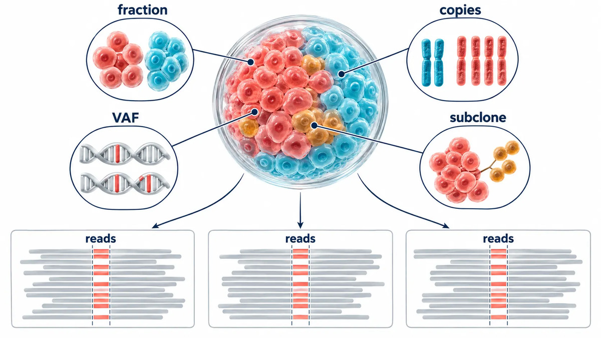 Figure
2: Variant allele frequency reflects tumor fraction, copy number, mutant-copy multiplicity, and clonal composition rather than purity alone.
Build the Pre-Sequencing Reference Package First
A purity call is strongest when the organoid is compared with an existing reference package rather than judged against a generic tumor profile. Before sequencing a new passage, collect the most decision-useful source information available: a pathology summary, baseline morphology images, known tumor-associated variants, a tissue or earlier-passage DNA profile, expected CNV landmarks, and any normal comparator. The package does not need to be exhaustive. It needs to contain at least one molecular feature that normal cells are unlikely to reproduce.
When a project has a defined driver or a small set of informative variants, an economical targeted assay may be the most efficient gate. When the variant history is incomplete, a broader DNA approach becomes more valuable. Gene Panel Sequencing Service options can support focused confirmation of known loci, whereas Whole Exome Sequencing may be more appropriate when several variants and broader CNV context are required. The choice should follow the evidence gap, not an automatic preference for the largest assay. If the question later expands from culture purity to tissue-to-model molecular concordance, use the separate multi-omics organoid fidelity framework rather than stretching this release gate beyond its purpose.
Define the release question before submitting material. For example: "Does passage 4 retain two baseline variants and the three largest chromosomal imbalances?" is a decision-ready question. "What mutations are in this organoid?" is not. The first can yield a PASS, HOLD, or FAIL decision. The second can invite a broad data-generating exercise with no predefined acceptance logic.
Gate 1: Use Morphology, Histology, and Protein Markers as a Rapid Screen
Bright-field images are useful for detecting a sudden shift in architecture, lumen formation, budding, compaction, debris, or heterogeneous colony size. They are not enough to label a culture as tumor-derived. Both normal and tumor epithelial populations can form cystic or solid 3D structures, and morphology can change with medium, matrix, and density. Use images to decide whether a well deserves investigation, not as the final release criterion.
Histology and marker assessment add spatial context. An organoid histology review can show architecture, epithelial organization, and the distribution of selected markers across the culture. The most useful markers are those selected from the original tissue knowledge and the project question, not a universal checklist. For instance, a lineage marker may confirm epithelial identity while a tumor-associated marker may help flag a population change. Neither should override DNA evidence if the two disagree.
Use Gate 1 as a triage screen with three possible outcomes. Stable morphology plus expected marker pattern supports progression to DNA confirmation. A sudden or inconsistent pattern triggers an immediate HOLD and comparison with earlier material. A clearly non-matching pattern may justify re-derivation without spending sequencing capacity. This gate is deliberately fast because it prevents sending an obviously uncertain culture into a deeper workflow.
Gate 2: Confirm Known Tumor Variants with a Focused DNA Assay
When baseline tissue or an early organoid passage contains a well-supported tumor-associated variant, targeted confirmation is often the most direct purity test. The goal is not to rediscover the entire genomic profile. It is to ask whether the expected molecular anchor remains detectable at a level consistent with the culture's intended use. A targeted locus assay can be designed around one or several high-confidence variants, ideally including more than one genomic region when material allows.
Interpret the result longitudinally. Compare the current VAF with the baseline tissue and, if available, an earlier organoid passage prepared with the same assay. A marked downward shift across multiple expected variants is more persuasive than a low value at one site. At the same time, do not set a universal VAF cutoff. A heterozygous driver in a copy-neutral clone behaves differently from an amplified allele or a subclonal variant. Read depth, local copy number, and possible allele dropout should be recorded alongside the VAF.
For cultures with limited DNA, the pre-analytical quality of the extract matters as much as the sequencing assay. Confirm concentration, fragmentation state, and extraction consistency before treating an absent call as biology. If a variant is absent in one low-input aliquot but present in another, the correct conclusion is HOLD, not "normal overgrowth confirmed." Carefully planned organoid exome sequencing can provide a broader resolution path when a small panel cannot settle the result.
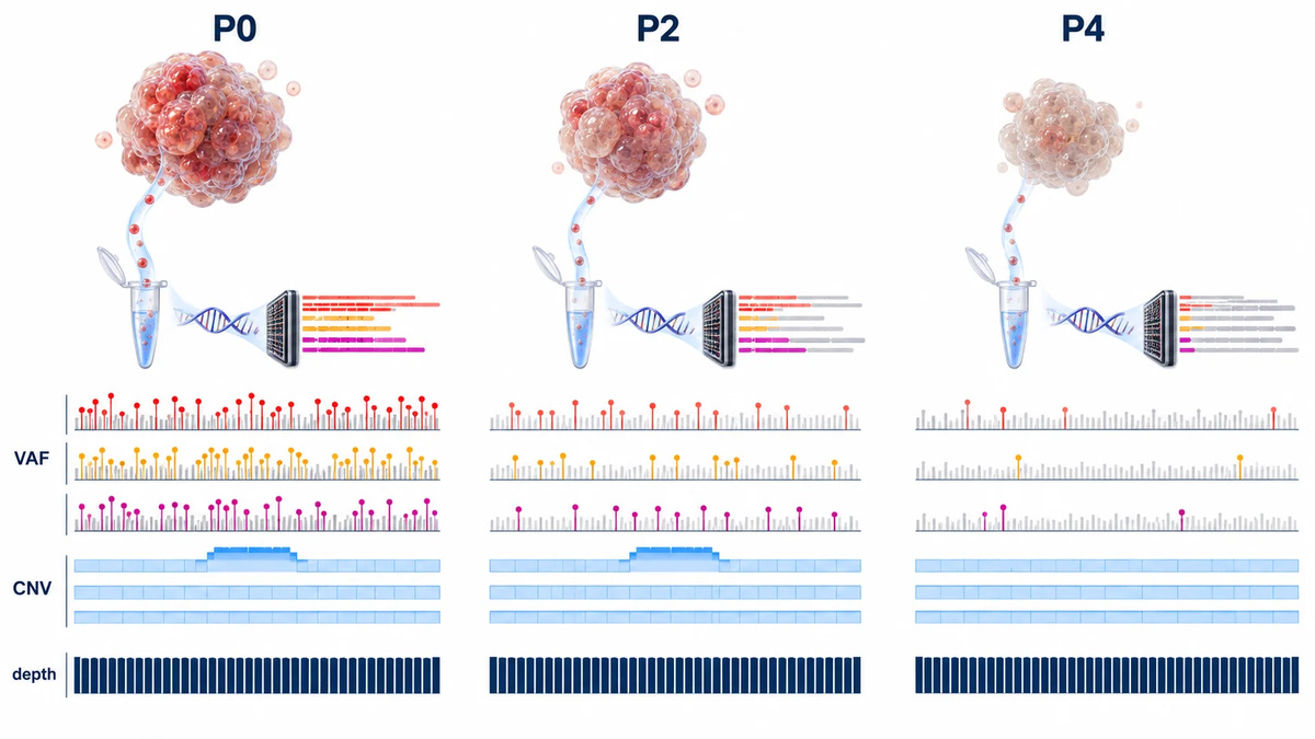 Figure
3: Targeted tumor variants should be evaluated as a longitudinal pattern with read depth and copy-number context, not as a single VAF cutoff.
Gate 3: Use CNV Profiles When No Single Driver Can Carry the Decision
Some cultures have no known dominant mutation, have multiple weakly informative variants, or come from diseases with diverse driver landscapes. In those settings, CNV structure may provide the most practical tumor-purity signal. The question is not whether every segment matches the source tissue exactly. It is whether the culture retains a recognizable pattern of broad or focal copy-number landmarks that distinguishes it from a normal diploid epithelial population.
Choose two or more informative landmarks from baseline material where possible: a broad arm-level gain, a recurring loss, and a focal event can form a useful composite signature. In the organoid, look for coordinated attenuation or disappearance of those events. A single smooth profile may reflect low DNA quality, poor normalization, or biological purity loss. A multi-locus pattern that moves toward diploidy is more concerning, especially when it agrees with falling driver VAFs.
CNV Sequencing Services can support this DNA-led gate, while a carefully documented variant calling workflow ensures that copy-number interpretation is not separated from variant evidence. Be cautious with absolute purity outputs from a single algorithm. Published mixture experiments show that computational purity estimates can vary materially between tools and may underestimate known tumor fractions at lower purity. Treat the numerical estimate as one part of the evidence package, not a stand-alone release certificate.
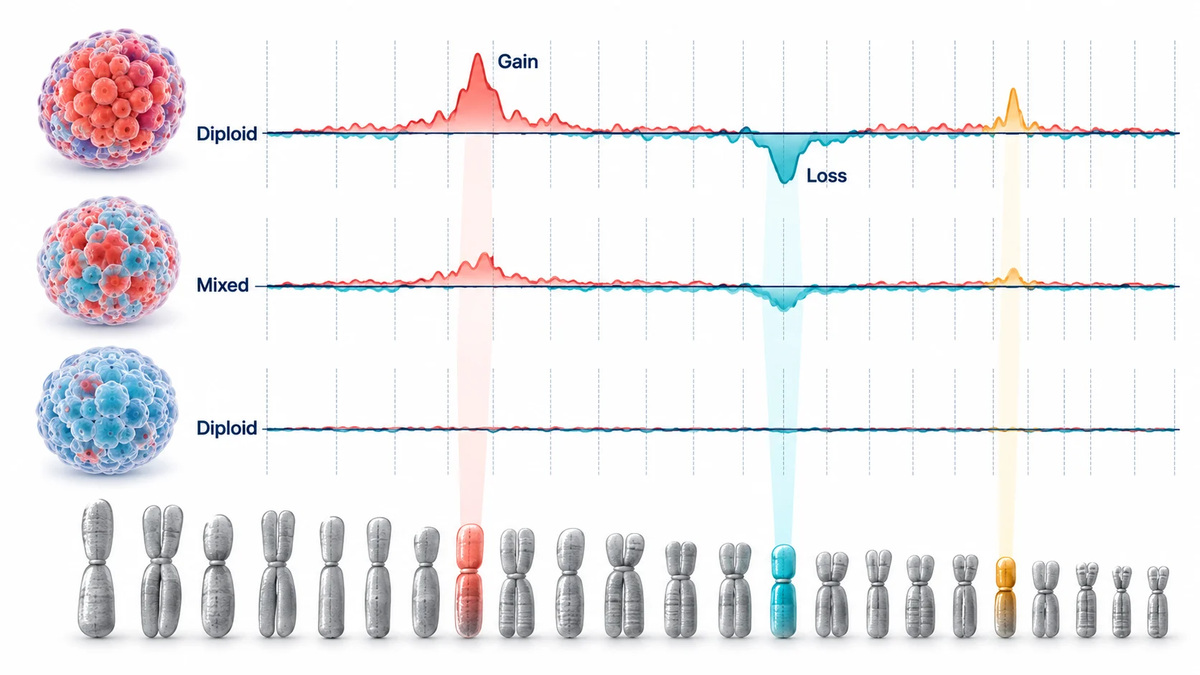 Figure
4: Normal-cell dilution flattens tumor copy-number gains and losses toward a diploid baseline, making multiple CNV landmarks more informative than a single segment.
Gate 4: Use RNA Evidence as Support, Not Final Proof
RNA data can reveal whether the culture has moved toward a normal epithelial differentiation program, a stress state, or a transcriptional profile inconsistent with the intended model. It is particularly useful when paired with reference RNA from earlier passages or source material. However, RNA should not be used alone to prove tumor purity. Culture itself can produce tumor-like proliferation and pathway signatures in genetically normal epithelial cells. Conversely, a true tumor population can alter expression after adaptation to matrix and medium.
Use transcriptomics to ask bounded questions. Does the current culture cluster with an earlier qualified passage? Are normal-lineage markers increasing while expected tumor-associated programs decline? Is the RNA change consistent with a DNA-based shift, or does it appear as an isolated transcriptional adaptation? A standard RNA-Seq comparison can answer these questions when RNA quantity is sufficient. For very small organoid pellets or defined micro-well samples, Ultra Low RNA Sequencing may preserve the ability to use RNA as a supporting layer.
If DNA and RNA disagree, the resolution should favor the DNA evidence for the narrow purity question. RNA disagreement is still valuable: it may reveal a state shift, medium effect, or emerging subpopulation that warrants a separate investigation. It is not a reason to override stable VAF and CNV evidence with an expression-only conclusion.
The Pre-Sequencing Tumor-Purity Decision Matrix
The most useful framework is not "run every assay on every sample." It is a tiered decision matrix that escalates only when the previous evidence is insufficient.
| Evidence question | Primary readout | What a concerning result looks like | Next action |
|---|---|---|---|
| Has the culture visibly changed? | Bright-field images and histology | Architecture or marker pattern changes across passages | HOLD; compare with early images and DNA baseline |
| Are known tumor anchors retained? | Targeted variants and VAF trend | Loss or coordinated VAF decline across expected variants | Check depth and copy number; escalate to CNV |
| Does the DNA retain tumor-specific structure? | CNV landmarks and allelic imbalance | Broad flattening toward diploidy or lost landmark pattern | HOLD or FAIL; re-derive or enrich where feasible |
| Does the cell state support the DNA finding? | RNA comparison | Shift toward normal lineage or discordant composition signal | Use as corroboration; investigate culture variables |
The matrix supports proportional effort. A culture with stable morphology, two retained variants, and stable CNV landmarks may be ready for the planned sequencing assay. A culture with no known driver and an ambiguous CNV profile should not be pushed through because it has already expanded. It needs a defined HOLD pathway. In projects that need a coordinated DNA/RNA decision rather than isolated files, organoid sequencing analysis can keep the comparison logic consistent across passages and evidence types.
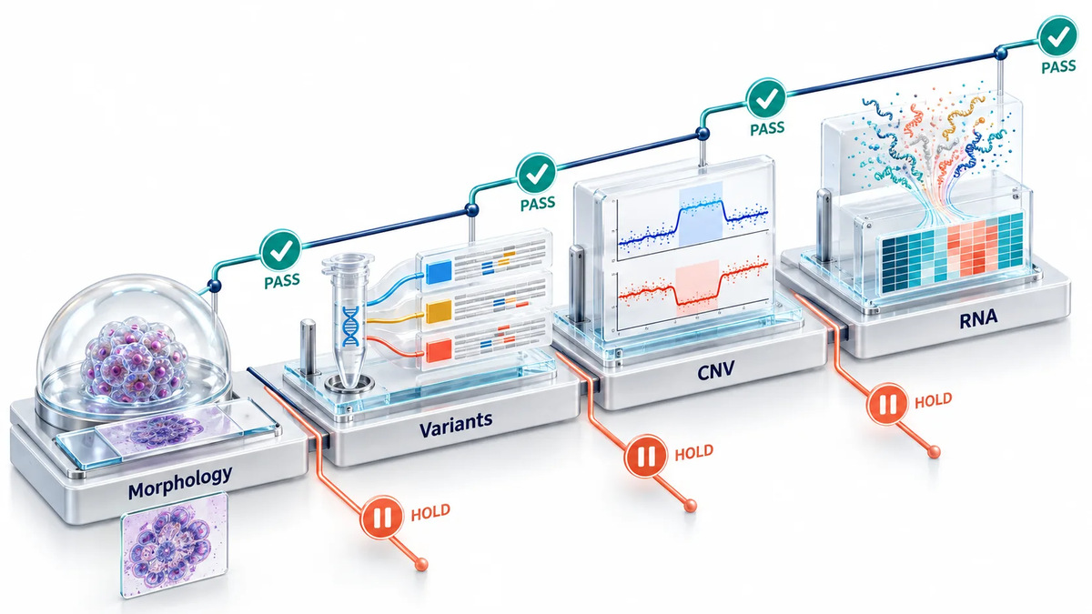 Figure
5: A pre-sequencing tumor-purity gate integrates morphology, targeted variants, CNV landmarks, and RNA support into a proportional escalation workflow.
PASS, HOLD, or FAIL: A Practical Release Rule
A pre-sequencing gate should end in an action, not a narrative. Define the action before samples are collected.
- PASS: morphology is not concerning; at least one DNA anchor is retained; and the CNV or multi-variant pattern is compatible with the qualified baseline. RNA, if used, does not contradict the DNA conclusion.
- HOLD: one evidence stream is inconclusive, DNA quality is inadequate, or morphology and molecular data disagree. Do not sequence at scale yet. Repeat the minimal decisive assay from a fresh aliquot or add the next DNA layer.
- FAIL: expected tumor anchors are absent or strongly diluted across a quality-controlled assay, CNV landmarks have collapsed toward a normal profile, and the result is supported by a second independent evidence stream.
When morphology or RNA appears reassuring but the variant and CNV evidence is inconsistent, do not convert the conflict into a PASS by averaging the signals. Assign HOLD. Re-test the smallest decisive DNA panel from a fresh, independently extracted aliquot, then compare the result with the same baseline anchors. This rule keeps a visually convincing culture or an adaptive expression state from overruling the evidence most directly linked to tumor-cell composition.
These labels are project rules, not universal biological thresholds. A culture intended for exploratory expression work may tolerate a different residual mixture than a culture intended for mutation-resolved comparison. The practical discipline is to write the purpose, minimum evidence, and action rule into the study brief before data are generated. This makes the decision reproducible across operators and batches.
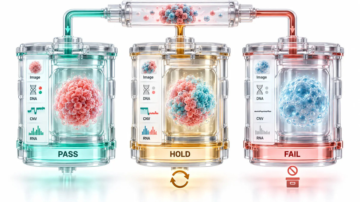 Figure
6: PASS, HOLD, and FAIL decisions should be predefined from convergent evidence rather than inferred after a large sequencing run.
What to Do When Normal-Cell Overgrowth Is Detected
A failed purity gate is not a reason to erase the record. It is a reason to preserve the chain of evidence and choose the most efficient recovery route. First, bank the available material with clear status labels rather than continuing to expand an unqualified culture. Then compare the culture log with a qualified reference: when did the VAF or CNV change appear, and what changed in medium, matrix, density, or handling?
Recovery options depend on the biological setting. An earlier banked passage may retain the desired population. Re-derivation from archived source tissue may be preferable to prolonged rescue. In some projects, manual selection of a distinct organoid morphology or single-organoid expansion can help separate subpopulations, but it must be followed by the same molecular gate. Adjusting the culture condition may reduce the selective advantage of normal cells, yet any change in medium or matrix should be treated as a new process variable and requalified.
When the culture is likely mixed rather than fully normal, do not overinterpret bulk averages. A small follow-up experiment may be enough to decide whether subclonal sampling is worthwhile or whether the model should be re-established. The appropriate sequencing scale is the one that answers this decision. A broader Organoids Sequencing Service can be planned after the purity gate has established that the material is fit for the intended research question.
Minimum Reporting Standard for a Tumor-Purity Gate
Purity decisions become reusable only when they are documented as a small, structured packet. Record the source and organoid passage, matrix and medium version, date of extraction, DNA and RNA input quality, baseline molecular anchors, observed VAFs with depth, CNV landmarks assessed, histology or marker summary, and the PASS/HOLD/FAIL outcome. Include the reason for a HOLD or FAIL and the next planned action.
This packet has two benefits. It gives later analysts the context needed to interpret a sequencing result. It also exposes recurring process failures. If several cultures lose expected variants after the same matrix change or split routine, the answer is no longer hidden in individual project folders. A disciplined organoid research solution can connect this sample history to the choice of molecular assay and downstream interpretation, while retaining the project's specific acceptance criteria.
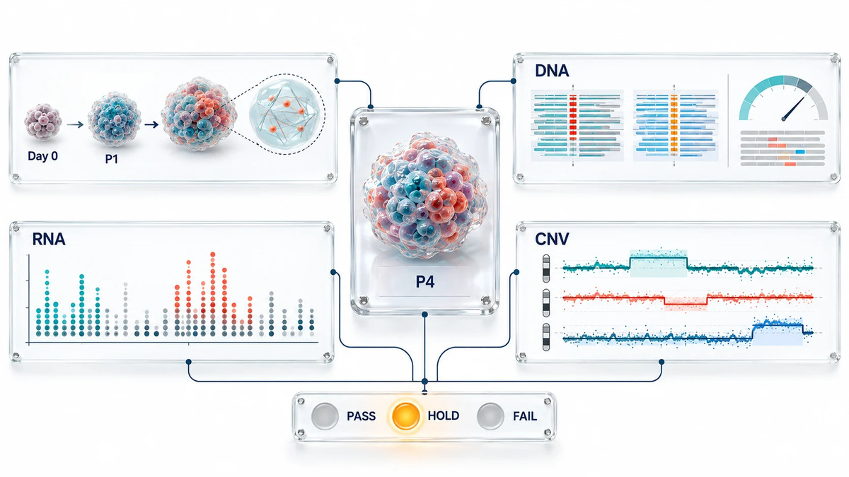 Figure
7: A concise tumor-purity release record links culture history, DNA anchors, CNV landmarks, RNA support, and the final action for each organoid passage.
Frequently Asked Questions
Can morphology alone confirm that an organoid culture is tumor-derived?
No. Morphology is a useful early warning signal, but normal and tumor epithelial populations can both form viable 3D structures. Use it to trigger molecular confirmation, not to replace it.
Is a missing tumor variant always evidence of normal-cell overgrowth?
No. Low input, coverage loss, allele dropout, a subclonal baseline variant, or incorrect reference material can also produce a missing call. Confirm assay quality and use a second independent DNA evidence stream.
What is the fastest molecular gate when baseline variants are known?
A focused multi-locus DNA assay is usually the most efficient first step. It should report VAF together with read depth and, where relevant, local copy-number context.
What if no known driver mutation is available?
Use a composite CNV signature from baseline material, ideally with more than one altered region. If that remains ambiguous, generate a broader DNA reference rather than relying on RNA similarity.
Can RNA-seq replace DNA confirmation?
No. Culture conditions can make normal organoid cells adopt tumor-like expression programs. RNA is best used to support, explain, or challenge a DNA-led conclusion.
Should every passage be sequenced?
Not necessarily. Define high-value checkpoints, such as early establishment, a major process change, and immediately before a high-value sequencing study. Use lower-cost focused gates between those points.
What should happen after a HOLD result?
Pause the planned large assay, repeat the minimal decisive test from a fresh aliquot, and compare with earlier qualified material. A HOLD is designed to prevent an irreversible choice made from incomplete evidence.
Can a mixed culture still be useful for research?
Possibly, but only if the mixture is measured and compatible with the stated research question. It should not be represented as a high-purity tumor organoid model without evidence.
References:
- Tbeileh N, Timmerman L, Mattis AN, et al. Metastatic colorectal adenocarcinoma tumor purity assessment from whole exome sequencing data. PLoS ONE. 2023;18(4):e0271354.
- Wang R, Mao Y, Wang W, et al. Systematic evaluation of colorectal cancer organoid system by single-cell RNA-Seq analysis. Genome Biology. 2022;23:106.
- Xu X, Kumari R, Zhou J, et al. A living biobank of matched pairs of patient-derived xenografts and organoids for cancer pharmacology. PLoS ONE. 2023;18(1):e0279821.
For research use only. Not for use in diagnostic procedures, clinical decision-making, patient stratification, therapeutic selection, or clinical trials.