

 Sample Submission Guidelines

CRISPR Off-Target Risk Is Cell- and Organ-Specific: A Preclinical Assay Design Framework
CRISPR off-target assessment is often reduced to a familiar sequence: predict similar genomic sites, sequence a panel, and report whether unexpected edits were detected. That sequence is useful, but it is not yet a preclinical assay design. It does not answer whether the tested cells represent the edited product, whether a whole-organ DNA extract has diluted a rare edited lineage, whether a structural rearrangement shares a cell with another lesion, or whether the chosen time point has already selected damaged cells out of the sample. A negative bulk result can therefore be informative without being universal.
A more defensible approach begins with the editing context. The same guide can behave differently across cell states, tissues, exposure durations, and repair environments. Recent single-cell and in vivo work has reinforced that individual cells can carry private off-target and translocation profiles, while bulk tissues can show organ-distinct editing and repair patterns. The practical implication is not that every project needs every possible assay. It is that bulk confirmation should be the backbone of the evidence package, with cell-resolved, structural-variant, and organ-context layers added when the biological risk question demands them.
This Resource presents a research-use framework for making those choices. It is designed for teams planning nuclease-based editing studies, multiplex edits, ex vivo cell products, or in vivo editing experiments that need to turn a long method list into a proportionate experimental plan.
Why a Bulk-Negative Result Is Not a Universal Safety Statement
Bulk targeted sequencing estimates the average frequency of a predefined event in the DNA molecules that enter a library. That is exactly why it is valuable: it can compare candidate sites across replicates, quantify recurring low-frequency indels, and provide a common analytical baseline across process conditions. However, it is also an average. A rare edited progenitor, a small subpopulation in a heterogeneous tissue, or a clone carrying two linked abnormalities may contribute too few molecules to change the bulk allele fraction appreciably.
Consider two samples with the same apparent 0.05% off-target frequency. In one, many cells each carry a single low-level event. In the other, a small clone carries the off-target edit, a large deletion, and a translocation together. Bulk frequency alone cannot distinguish those situations. The biological questions, and the sensible follow-up assays, are different. The first may call for more replicate and time-point data; the second may justify cell-resolved characterization and structural-variant confirmation.
Detection is also bounded by physical inputs. Read depth is not the same as sensitivity. Meaningful sensitivity depends on the number of genome equivalents recovered, library conversion efficiency, the number of independent molecules after deduplication, sequencing error background, and the unedited control distribution. A report should therefore avoid the phrase "no off-target effects." A more accurate statement is: no event above the prespecified detection boundary was observed at the nominated sites in the tested sample type, at the stated input and coverage.
 Figure 1: How bulk averaging can conceal cell-private CRISPR outcomes.
What Cell- and Organ-Context Studies Change—and What They Do Not
Cell-resolved studies show that bulk assays cannot identify whether distinct edit outcomes co-occur within the same cells, while in vivo off-target frameworks treat tissue exposure and cell type as explicit sampling variables rather than inferring them from a single tissue result.[2,4] The practical value is not a universal organ ranking. It is a design rule: use organ- and cell-context information to decide whether bulk data are sufficient for a defined exposure model.
The boundaries matter just as much. Whole-organ measurements may be dominated by the most abundant cell type, and organ-level differences can reflect cell composition, delivery exposure, repair state, and differential survival of damaged cells. The results support context-matched escalation layers for higher-risk studies; they do not establish a universal pass/fail threshold.
This distinction improves planning. Do not ask whether bulk testing is "enough" in the abstract. Ask whether the current experiment could plausibly hide a consequential event because of lineage dilution, multiplex cleavage, persistent editor exposure, a self-renewing cell population, or broad biodistribution. If the answer is yes, define the escalation step before samples are generated.
Start with Five Design Variables, Not with an Assay Name
An assay plan should be written after five variables are specified. First, define the editor mechanism: a double-strand-break nuclease, base editor, prime editor, or another system does not create the same event spectrum. Second, define delivery and exposure: transient RNP, mRNA-lipid nanoparticle delivery, and persistent vector expression create different windows for cleavage and repair. Third, define the biological context: the final edited cell population, target tissue, and any non-target tissues where the editing system may be detected. Fourth, define event classes of concern: substitutions, indels, large deletions, inversions, copy-number changes, translocations, or vector integration. Finally, define the analytical question: candidate-site discovery, quantitative confirmation, co-occurrence in individual cells, persistence, or genome-wide structural characterization.
These variables prevent a common failure mode: using a technically sophisticated assay that answers the wrong question. A biochemical discovery method can nominate many candidate sites, but it does not prove those sites are edited in the relevant cell type. A deep amplicon panel can quantify known loci, but it cannot establish that all important loci were nominated. A bait-based translocation assay can discover rearrangements anchored at a selected break, but it may miss off-target-to-off-target products. The appropriate study is an evidence stack, not a contest to select one "best" assay.
 Figure 2: Five variables that define a context-matched CRISPR off-target study.
Layer 1: Build a Candidate-Site Nomination Stack
Candidate nomination should start with sequence-aware computation but should not end there. Guide similarity, PAM compatibility, mismatch position, bulges, and genomic annotation provide a rational first list. For studies involving diverse donors or disease-associated haplotypes, variant-aware analysis is especially important. A variant can create a PAM, reduce the effective mismatch count, or change the local sequence context of a site that a reference-genome-only screen would rank as low concern.
Experimental nomination adds evidence that a guide can cut beyond its intended locus. Cell-free methods can be sensitive and scalable, while cell-based or in situ approaches preserve more of the intracellular context. Their outputs should not be treated as interchangeable. Cell-free methods can over-nominate accessible DNA fragments that would not be reached in a given cell, whereas cell-based methods can miss events when delivery, tag incorporation, or repair-state requirements are not met. For that reason, a practical candidate list often combines computational prediction with one or more empirical nomination methods rather than relying on their intersection alone.
Filtering should be predefined and transparent. Frequency, reproducibility, guide similarity, genomic context, and evidence across methods can all inform prioritization. An overly stringent sequence-distance cutoff may remove true sites, while an unranked list may make targeted confirmation inefficient. The goal is neither the longest panel nor the shortest panel. It is a risk-tiered panel that preserves plausible sites while reserving additional assays for loci with higher biological consequence.
For projects that require a targeted confirmation panel after discovery, CRISPR Off-Target Validation can provide a direct route from nominated loci to sequencing-based verification. Early design of primer compatibility and locus spacing also makes later translocation follow-up more feasible.
Layer 2: Confirm Events in the Cells That Matter
Confirmation should use edited material that represents the intended biological context as closely as practical. A convenient immortalized line can support method development, but it should not be the sole evidence source when the final project involves primary cells, differentiated derivatives, or a complex in vivo target. An April 2026 FDA draft guidance for nonclinical NGS studies supporting IND/BLA submissions for investigational human genome-editing products similarly highlights representative cell types, comparable on-target editing conditions, biological replicates, and biodistribution-informed tissue selection.[5] It is non-binding and is cited here only as a source of research-design considerations, not as regulatory advice.
Multiplexed amplicon panels work well when the candidate list is defined and the key question is per-site frequency. A robust panel includes unedited, process-matched controls; independently prepared biological replicates; on-target measurements; site-level coverage criteria; and a documented treatment of low-level background. Targeted Region Sequencing is appropriate when the study needs focused coverage across a risk-tiered locus list, while Amplicon Sequencing Services can support higher-throughput follow-up of defined PCR-compatible loci.
The confirmation design should include an input-genome budget. Before sequencing, specify the usable DNA mass or cell number, expected molecular recovery, minimum effective coverage per site, and the decision rule for a site that is detected in only one replicate. This makes a negative result interpretable. It also prevents the team from making a false comparison between a high-read library with few original templates and a moderately sequenced library with a much larger independent molecular input.
Layer 3: Add Structural-Variant and Translocation Evidence When the Edit Creates That Risk
Small indels do not exhaust the possible outcomes of genome editing. Double-strand breaks can also be associated with large deletions, inversions, copy-number changes, chromosomal translocations, or loss of primer-binding sites that make a short amplicon appear deceptively clean.[3] These events are not guaranteed, and their relevance depends on the locus, editor, cell state, and number of simultaneous cuts. But they should be considered explicitly when a project uses a nuclease, makes multiple edits, or operates in cells with long-term proliferative potential.
Baited translocation assays are useful because they enrich products that join a selected break to other loci. Their limitation is structural: an assay anchored only at the intended locus may underrepresent events between two off-target breaks. Targeted sequencing may also miss unbalanced products or events that remove a primer-binding site. The appropriate response is not to discard baited methods; it is to define their blind spots and add orthogonal confirmation when a signal appears.
Longer read designs can help resolve complex products spanning a candidate locus and its flanking sequence. Nanopore Target Sequencing is useful when a targeted long-read follow-up is needed to clarify large indels, inversions, or complex junction architecture. For a broader copy-number question, CNV Sequencing Services may be a better escalation layer than additional short-amplicon depth alone.
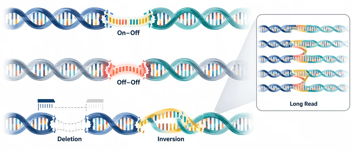 Figure 3: Translocation topologies and common targeted-assay blind spots.
When Single-Cell Resolution Changes the Answer
Single-cell analysis is most valuable when the unanswered question is about co-occurrence, clonality, or rare subpopulations. It can reveal whether an off-target edit and a structural event are present in the same cell, whether different clones carry different combinations of edits, and whether a small high-concern population is diluted in bulk DNA.[4] It is especially relevant for multiplexed edits, self-renewing stem or progenitor populations, edited immune-cell products, and projects where individual clones may expand after editing.
Single-cell does not mean unbiased genome-wide discovery by default. A targeted single-cell panel still depends on which sites were nominated. Clonal expansion can increase the detectability of a private event but can also select against cells with impaired fitness. Direct single-cell amplification avoids some expansion bias but introduces allelic dropout and amplification artefacts. A sound plan states which trade-off is being accepted and uses bulk data to retain population-level frequency context.
A useful operational trigger is this: add single-cell analysis when a bulk signal is low-frequency but repeated, when several double-strand breaks are introduced together, when a lineage has high expansion potential, or when a project decision depends on whether events co-occur in the same cells. Do not add it merely because higher resolution sounds more complete.
Translate Biodistribution into an Organ Sampling Plan
For in vivo editing, tissue selection should not be an arbitrary list of major organs. Begin with the intended target tissue. Then add tissues or organs with demonstrated product exposure, tissues with relevant cell biology, and any site where an observed signal would materially change the risk interpretation. This turns biodistribution from a separate workstream into an input for genome-integrity analysis.
Whole-organ DNA is often the sensible first layer because it offers broad coverage and straightforward comparison. But a whole-organ result can be dominated by unedited cells. If the delivered editor is expected to reach a defined minority lineage, consider cell enrichment, sorting, or a parallel marker-based readout. The purpose is not to manufacture a cell-type signal. It is to determine whether the sample actually has the analytical power to address the biological question.
Where RNA or chromatin context is needed to interpret candidate-site accessibility, pair genome editing data with appropriately scoped ATAC-Seq or RNA-Seq data. These layers can generate hypotheses about accessibility, cell-state composition, and repair context. They should not be used to claim that an open chromatin peak or expression value caused a particular off-target event without direct perturbation evidence.
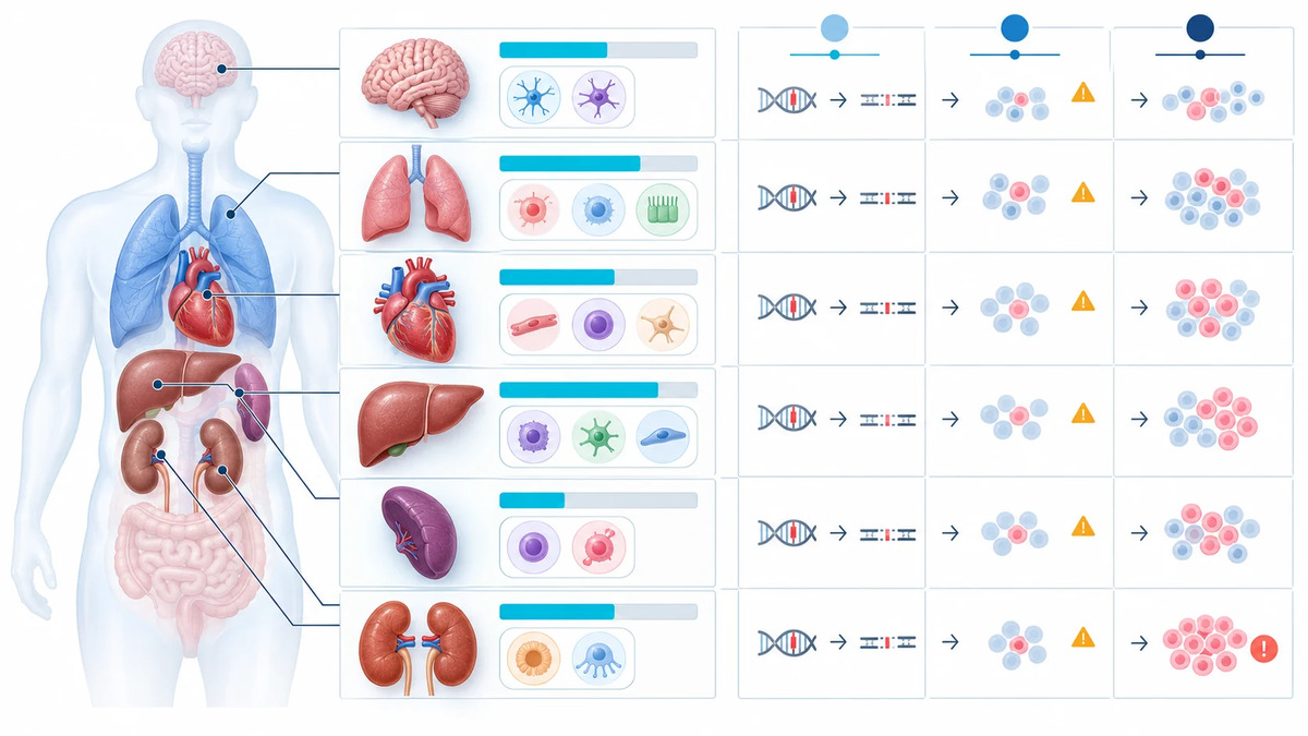 Figure 4: Organ and cell-type sampling matrix guided by delivery exposure and risk.
Plan Early, Intermediate, and Late Time Points
Time is an analytical variable, not a procedural afterthought. An early collection can capture initial cleavage and repair before damaged cells are lost. An intermediate collection can measure stabilized indels and rearrangements after the acute repair window. A later collection can test whether a signal persists, disappears through negative selection, or becomes enriched through clonal growth. For persistent delivery systems, later sampling may also be required to evaluate the consequence of extended editor exposure.
The correct schedule depends on the delivery format and tissue turnover. A short-lived RNP experiment in cultured cells may need closely spaced early and post-recovery collections. A vector-based in vivo study may need a wider window that separates exposure, repair, and persistence. Predefining the rationale prevents a late low-frequency result from being misread as evidence that an early event never occurred.
A Risk-Triggered Assay Architecture
| Editing context | Core evidence | Escalate when |
|---|---|---|
| Single, transient ex vivo edit | Nomination plus bulk candidate-site confirmation | Recurring low-frequency signal or high-concern locus |
| Multiplex ex vivo editing | Bulk confirmation plus structural-variant assessment | Multiple DSBs, rearrangement signal, or expanded-cell product |
| Stem or progenitor cell editing | Context-matched bulk panel and persistence time point | Potential clonal expansion or co-occurring abnormalities |
| In vivo targeted delivery | Target-tissue confirmation and biodistribution-informed organ panel | Non-target exposure, heterogeneous tissue, or prolonged expression |
| Persistent vector-mediated editing | Repeated bulk assessment and structural-integrity plan | Vector integration question, delayed editing, or organ-specific signal |
The value of this architecture is proportionality. A single transient edit in a limited ex vivo context does not automatically require a multi-organ, single-cell program. Conversely, a broad-exposure or multiplex project should not rely on a single convenient cell line and a one-time amplicon panel. A written escalation rule makes the plan defensible before data are seen.
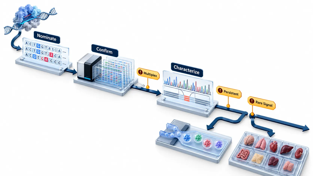 Figure 5: Risk-triggered escalation from nomination to organ-resolved and single-cell evidence.
Minimum Controls and Reporting Elements
At minimum, preserve an unedited control, a delivery or process-matched control where relevant, biological replicates, and on-target editing measurements. Document how candidate sites were nominated, how panels were filtered, what material entered the library, and what coverage remained after quality control. For translocation work, state which junction classes the assay can observe and which products may be missed. For tissue studies, record tissue composition, enrichment strategy, and the basis for organ selection.
Interpretation should remain evidence-bounded. A detected event may warrant confirmation, frequency estimation, persistence testing, and genomic-context review. It does not by itself establish functional consequence. Conversely, a low-frequency event should not be dismissed solely because it is below a generic threshold if it is reproducible, structurally complex, enriched over time, or located in a high-concern region. The study design should specify how those facts change the next experiment.
For broader genome-level follow-up rather than a candidate-site panel, Whole Genome Sequencing can serve as one component of a structural and variant-context strategy. If viral delivery is part of the exposure model, AAV Integration Site Analysis should be considered as a distinct question from nuclease off-target confirmation.
Match Each Assay to Its Blind Spot Before You Generate Samples
Every assay leaves an evidence gap. Computational tools may miss a donor-specific sequence that creates a new compatible site. Cell-free discovery can nominate sites that are not cut in the final cell type. Cell-based discovery can be limited by delivery efficiency, transient repair signals, or the need for an inserted tag. A targeted panel cannot discover a locus outside the panel. Long reads provide breakpoint context but do not automatically deliver the molecule counts needed for precise low-frequency quantification. Single-cell analysis resolves co-occurrence but trades breadth, input requirements, and cost against cell-level detail.
The practical answer is not to stack assays indiscriminately. Create a one-page blind-spot register before the first pilot. For every method, record the event class it can observe, the sample it requires, the lower-bound sensitivity that can be supported by available input, and the failure mode that requires an orthogonal method. For example, if a multiplex nuclease experiment uses short amplicons for routine frequency measurement, designate a subset of higher-risk loci for long-range or long-read validation. If a cell-based nomination assay uses a surrogate line, specify how the highest-ranked sites will be retested in primary or product-relevant cells. If whole tissue is used, predefine the tissue composition information needed to determine whether an enriched-cell experiment is warranted.
This exercise also prevents an unhelpful interpretation of method disagreement. A site nominated by a biochemical assay but absent in a product-relevant cell sample is not automatically a false positive; it may simply lack access or exposure in that tested context. Conversely, an event detected in relevant cells but missed during nomination may indicate a panel design gap, an individual genetic variant, or a context-specific repair outcome. The right response is to trace the evidence boundary, not to decide that one platform "won."
Use Functional and Multi-Omic Readouts as Context, Not as a Shortcut to Causality
ATAC-seq, RNA-seq, methylation profiling, and cell-surface phenotyping can add useful biological context to an off-target program. They can help determine whether an edited tissue contains the cell population of interest, whether a candidate locus sits in an accessible region, or whether an observed clone has a distinct transcriptional state. They can also guide sample enrichment and help separate a tissue-composition shift from a genuine change in editing outcome.
These measurements must not be overinterpreted. Open chromatin is not proof that a site will be cut, and a transcript-level difference is not proof that a low-frequency edit caused the observed phenotype. The strongest use case is hypothesis prioritization: combine editing calls with context data, formulate a specific follow-up question, then test it with a matched control or orthogonal assay. Teams needing to jointly organize such layers can use a defined Multi-Omics Service strategy to keep sample identity, time points, and data integration rules aligned from the start.
FAQ
Can deep bulk sequencing replace single-cell analysis?
No. Deep bulk sequencing improves population-level sensitivity at nominated sites, but it does not show whether multiple events coexist in the same cell or whether a rare clone is driving the signal.
Should every CRISPR project include organ-specific profiling?
No. It is most relevant for in vivo editing when target biology, biodistribution, delivery persistence, or non-target exposure makes tissue context important.
How many candidate off-target sites belong in a confirmation panel?
The panel should be risk-tiered rather than fixed in size. Include sites supported by empirical nomination, high-concern sequence matches, variant-created candidates, and loci whose genomic context would change interpretation.
When is long-read confirmation useful?
Use it when short amplicons cannot resolve large indels, inversions, complex junctions, or suspected primer-site loss around a candidate locus.
Does open chromatin prove that an off-target site will be edited?
No. Accessibility may help explain context, but sequence compatibility, delivery, nuclease exposure, repair state, and cell composition also influence the measured outcome.
How should a negative result be reported?
State the tested sample type, nominated sites, effective molecular input, coverage criteria, controls, and prespecified detection boundary. Avoid presenting a negative result as proof of universal absence.
For research use only.
References:
- Guo C, Ma X, Gao F, Guo Y. Off-target effects in CRISPR/Cas9 gene editing. Frontiers in Bioengineering and Biotechnology. 2023;11:1143157. doi:10.3389/fbioe.2023.1143157.
- Wienert B, Cromer MK. CRISPR nuclease off-target activity and mitigation strategies. Frontiers in Genome Editing. 2022;4:1050507. doi:10.3389/fgeed.2022.1050507.
- Aussel C, Cathomen T, Fuster-García C. The hidden risks of CRISPR/Cas: structural variations and genome integrity. Nature Communications. 2025;16:7208. doi:10.1038/s41467-025-62606-z.
- Kalter N, Gulati S, Rosenberg M, et al. Precise measurement of CRISPR genome editing outcomes through single-cell DNA sequencing. Molecular Therapy—Methods & Clinical Development. 2025;33(2):101449. doi:10.1016/j.omtm.2025.101449.
- U.S. Food and Drug Administration. Safety Assessment of Genome Editing in Human Gene Therapy Products Using Next-Generation Sequencing. Draft Guidance for Industry; April 2026. Docket FDA-2026-D-1255.