
 Sample Submission Guidelines
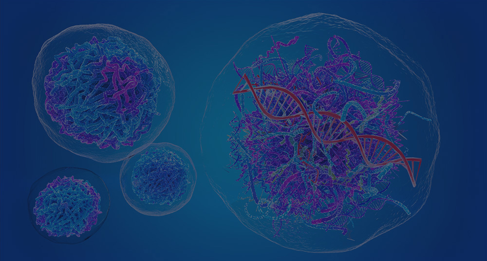
Meta Intent: Provide a rigorous structural biology perspective on how DNA compaction dictates gene accessibility, genome stability, and cellular identity.
Chromatin and chromosomes are often explained as if they were separate biological entities. In living cells, they are better understood as different physical and functional states of the same genome. Chromatin is the active working form of nuclear DNA. It is dynamic, locally accessible, and continually remodeled. Chromosomes are the highly condensed, segregation-ready form of that same material. They emerge when the cell must package DNA for inheritance rather than keep it open for ongoing transcription, replication, and repair.
This difference is not cosmetic. The physical state of DNA changes what the genome can do. It changes whether transcription factors can sample regulatory motifs, whether enhancers can contact the right promoters, whether repair enzymes can access lesions efficiently, and whether sister chromatids can separate cleanly during mitosis. In other words, genome folding is not a passive consequence of packing. It is a regulatory layer and a mechanical layer at the same time.
A useful framing is this: chromatin manages biological options, while chromosomes enforce biological order. During interphase, the genome must remain searchable. During mitosis, it must become transportable. These are different engineering problems. The cell solves them by moving DNA across a continuum of compaction states rather than keeping it in one permanent architecture. That continuum begins with nucleosomes, scales up through loops and compartments, and culminates in mitotic chromosomes that are compact enough to segregate but not so adhesive that they tear, bridge, or mis-segregate.
That is why chromatin biology now overlaps with spatial epigenomics, developmental biology, and cancer genomics. The key question is no longer simply what DNA sequence is present. The key question is how that sequence is physically deployed in space and time.
The Continuum of Compaction
Nucleosomes are the first control valve
The nucleosome is the fundamental repeating unit of chromatin. Around 147 base pairs of DNA wrap around a histone octamer composed of H2A, H2B, H3, and H4. This is often described as the first level of DNA packaging. That description is correct, but incomplete. Nucleosomes do more than shorten DNA. They determine which DNA surfaces are exposed, which are hidden, and how easily proteins can compete for access.
That means nucleosomes are not just structural beads. They are regulatory gates. A promoter may contain an ideal transcription factor motif and still remain silent if a stable nucleosome blocks the relevant binding surface. The opposite can also happen. A modest motif can become functional if nucleosome sliding, eviction, or histone-tail modification exposes it. Packaging and regulation therefore begin at the same place.
This principle helps explain why accessibility often changes before strong transcriptional changes become obvious. During lineage commitment, inflammation, or environmental stress, one of the earliest measurable events is often a local shift in nucleosome positioning or occupancy. The door opens before the room is used. That is why chromatin accessibility is so informative: it often marks the earliest reduction in regulatory resistance.
The nucleosome is also where chemical information begins to accumulate. Histone tails extend outward and can be acetylated, methylated, phosphorylated, ubiquitinated, or otherwise modified. These changes do not float above chromatin as abstract signals. They alter internucleosomal interaction, reader recruitment, local charge environment, and remodeler behavior. From the first level of organization onward, the genome is already being told how to behave.
Higher-order folding is real, but it is not uniformly textbook-like
For many years, genome compaction was drawn as a neat ladder: beads-on-a-string, then a regular 30-nm fiber, then larger coils, and finally metaphase chromosomes. That model was pedagogically useful, but native chromatin is more irregular. In cells, nucleosome arrays can adopt variable local configurations depending on transcriptional state, ionic environment, chromatin-binding proteins, and cell-cycle stage. Higher-order folding exists, but it is less uniform than older textbook diagrams suggested.
This matters because different compact states do not produce the same biological result. A region can be locally compact yet still remain remodeler-compatible. Another region can appear open in one assay but still be topologically constrained from its preferred regulatory partners. A third region can look dense in bulk data yet remain highly dynamic at short scale. The key variable is therefore not compaction alone. It is compaction plus mobility, compaction plus topology, and compaction plus biochemical context.
A useful case comes from nucleosome remodeling work on condensed arrays. Rather than treating condensed chromatin as a dead state, recent studies show that some compacted arrays can still support ATP-dependent nucleosome movement. That observation changes the way compaction should be discussed. A region is not defined only by how tightly it is packed. It is also defined by whether that packing remains internally adjustable.
This shift also helps explain why chromatin can support both stability and flexibility. Cells need enough order to avoid random polymer behavior, yet enough local plasticity to rewire transcription during development, stress response, and lineage restriction. If chromatin were too loose, enhancer choice would become noisy and genome integrity would suffer. If it were too rigid, developmental transitions would stall. The nucleus operates between those extremes.

Figure 1. Hierarchical compaction roadmap. A staged schematic showing how the eukaryotic genome progresses from DNA double helix to nucleosome, beads-on-a-string chromatin, folded chromatin fiber, looped higher-order domains, and a condensed metaphase chromosome.
Chromatin becomes chromosomes when the physical task changes
The most useful difference between chromatin and chromosomes is function. Interphase chromatin is optimized for regulated access. Mitotic chromosomes are optimized for mechanical fidelity. During interphase, the genome must support transcription, replication, repair, and long-range regulatory communication. During mitosis, those priorities shift. DNA must be condensed into individualized units that can resist spindle force, minimize entanglement, and segregate accurately.
This transformation is not a generic tightening event. It involves condensin complexes, topoisomerase II, histone-tail-mediated interactions, and large-scale loop restructuring. The goal is to generate chromosomes that are compact enough to move as discrete bodies, but not so sticky or improperly resolved that bridges form during anaphase. Mechanical robustness becomes the dominant requirement.
This perspective also explains why chromosome defects often have earlier chromatin causes. Replication stress, unresolved catenation, mistimed condensation, and abnormal chromatin stickiness can all compromise chromosome individualization later. What appears to be a mitotic failure may actually begin as a chromatin management problem several hours earlier in the cell cycle.
Another way to state the same principle is this: interphase chromatin is built for regulated search, while mitotic chromosomes are built for safe transport. One state must support selective contact. The other must support faithful partitioning. The genome cannot maximize both tasks at once, so it transitions between states with strong temporal control.
Functional Topology: TADs, Loops, and Nuclear Compartments
TADs reduce regulatory ambiguity
One of the major advances in 3D genomics was the realization that the genome is partitioned into recurring topological neighborhoods. Topologically associating domains, or TADs, are regions within which chromatin interactions are enriched relative to surrounding sequence. Their biological value is straightforward: they reduce regulatory ambiguity.
Enhancers do not search the entire chromosome arm equally. They preferentially sample a constrained neighborhood. Promoters are therefore exposed to a filtered contact environment rather than to every distal element that lies nearby in linear sequence. This improves regulatory specificity and reduces noise.
This principle becomes especially important during development. Many developmental defects can be understood not as sequence failure, but as neighborhood failure. A promoter may be intact. An enhancer may be intact. Yet a boundary defect can place them into the wrong topological relationship. Once the wrong regulatory partner becomes reachable, misexpression can follow even without coding mutation.
TADs are therefore best understood as structures that bias communication rather than as rigid boxes. They do not prevent every cross-boundary interaction. They make some contacts more likely and others less likely. In living systems, that probabilistic control is often enough to preserve cell-type-specific expression programs.
The CTCF-cohesin mechanism makes loop extrusion biologically useful
The strongest model for many local loops is cohesin-mediated loop extrusion constrained by CTCF. In this model, cohesin loads onto chromatin and reels in DNA until it encounters boundary elements, often CTCF sites in convergent orientation. This produces a looped domain with elevated internal contact probability.
The beauty of this mechanism is that it links structure to regulatory kinetics. A promoter does not need permanent contact with an enhancer. It needs contact to become likely enough, often enough, to support productive regulation. Loop extrusion changes that likelihood. It alters the frequency with which certain elements encounter each other and reduces the chance that others do.
A classic example of the power of local topology came from high-resolution contact mapping in human cells. When deeper maps became available, many more loops emerged than earlier studies could see, especially around promoter-rich regions. This did not simply add decorative detail to 3D maps. It changed how regulatory specificity was interpreted. The genome was not folded into a few coarse domains. It contained far more short-range structure than previously appreciated.
This is why changes in loop architecture often have outsized functional effects. A modest shift in anchor strength can alter promoter choice. A boundary inversion can redirect enhancer action. A reduction in cohesin residence time can soften insulation enough to matter. These are subtle physical changes, but they can produce decisive regulatory outcomes.

Figure 2. TAD formation and loop extrusion. A stepwise mechanism diagram illustrating how cohesin loads onto chromatin, extrudes a loop, and stabilizes a TAD-like domain when convergent CTCF boundary elements are encountered.
A/B compartments organize the nucleus at broader scale
TADs explain local organization. A/B compartments explain broader nuclear sorting. A compartments generally align with more active, gene-rich, and accessible chromatin. B compartments tend to align with denser, less active, and often lamina-associated chromatin. This larger-scale partitioning helps explain how cells with identical genomes can stabilize very different transcriptional programs.
A locus is regulated not only by its local loop environment, but also by the broader biochemical neighborhood in which it resides. A promoter located in a permissive interior environment behaves differently from one embedded in a repressive peripheral neighborhood. Compartmentalization does not dictate every transcriptional event, but it biases the landscape in which those events occur.
Compartment switching during differentiation illustrates this well. A locus may move away from a peripheral repressive region, gain local accessibility, acquire activating histone marks, and only then show strong transcription. The transition is often gradual. The locus first changes where it lives, then changes how it behaves. This is one reason spatial epigenomics has become so valuable. It connects chromatin state to nuclear geography rather than treating them as separate layers.
Compartment state also interacts with replication timing and repair environment. Early-replicating regions are often aligned with active compartments. Late-replicating regions more often align with repressive neighborhoods. This means compartmentalization influences not only transcription, but also the biochemical timing in which DNA transactions occur.

Figure 3. A/B compartment spatial map. A semi-flat nuclear organization diagram showing transcriptionally permissive A compartments enriched toward the interior and more compact B compartments preferentially positioned near the nuclear periphery and lamina.
Hi-C and Micro-C changed what local structure means
Hi-C made genome-wide chromatin contact mapping possible. It revealed chromosome territories, compartments, and large-scale folding principles. But Hi-C is limited by fragmentation strategy and effective resolution. Micro-C sharpened the picture by using nuclease digestion to capture finer-scale architecture, often approaching nucleosome resolution.
That difference matters because many biologically important decisions happen locally. Fine boundaries, short-range loops, promoter-adjacent structures, and subtle insulation effects can be blurred in standard Hi-C. Micro-C resolves more of this local organization. It is not just a technical upgrade. It changes what can be asked.
A clear case is the detection of short-range enhancer-promoter contacts. In lower-resolution maps, these interactions may merge into broader domains. In finer maps, they emerge as discrete structural features. This is particularly important when researchers want to connect chromatin architecture to precise regulatory output rather than to broad compartment trends.
The field is now moving from bulk maps to single-cell and multimodal maps. That shift is significant because a clean domain in bulk data may actually reflect several distinct sub-states across cells. Once architecture is measured cell by cell, genome folding becomes a cell-state trait rather than just a population average.
High-resolution methods also changed interpretation of boundaries. Some boundaries that seemed simple at coarse scale resolve into clusters of local insulation features, promoter-proximal loops, or nested interaction units. That finer picture is especially relevant for disease mechanism studies, where modest structural variation can rewire local architecture without dramatically altering large-scale compartment pattern.

Figure 4. Hi-C versus Micro-C technology comparison. A dual-column methods schematic contrasting workflow logic and output resolution, highlighting broader interaction patterns in Hi-C and finer local chromatin structure in Micro-C.
Chromatin Accessibility as a Regulatory Switch
Euchromatin and heterochromatin are dynamic modes
The classical distinction between euchromatin and heterochromatin remains useful, but it should be described dynamically. Euchromatin is generally more accessible and more transcription-permissive. Heterochromatin is generally denser and more restrictive. Yet these are not fixed materials. They are operating modes that can shift with signaling input, cell-cycle stage, developmental context, and environmental stress.
A region can become more accessible without immediately becoming highly expressed. A compact region can still be sampled by pioneer factors. A locus can remain low-output yet poised for activation later. Accessibility is therefore best understood as a change in permission, not a guarantee of transcription.
This distinction helps avoid one of the most common interpretation errors in epigenomics: treating open chromatin as proof of full activity. Open chromatin lowers the barrier to binding, but transcription still depends on cofactor availability, polymerase control, promoter competence, and appropriate 3D contact logic.
It is also important to remember that “closed” does not always mean inert. Some loci cycle between accessible and inaccessible sub-states. Others remain partially permissive to select classes of factors. In developmental systems, this graded behavior is common. Cells often prepare genes for future use by lowering the barrier before they fully commit to transcription.
Phase separation is relevant, but it is not a universal answer
Phase separation has become an important concept in nuclear biology because many chromatin-associated proteins contain intrinsically disordered regions and can form condensate-like assemblies. These assemblies may enrich specific regulators, stabilize repressive states, or alter local chromatin concentration. However, phase behavior should be treated as one organizing principle among several.
That nuance matters. Nuclear organization is also shaped by polymer behavior, loop extrusion, lamina interaction, sequence-specific recruitment, and chromatin modification. In some contexts, phase separation adds a useful explanatory layer. In others, it risks becoming a vague substitute for mechanism. Good technical writing keeps the idea precise: phase behavior can help define certain chromatin microenvironments, but it does not explain every feature of 3D genome architecture.
A balanced view is especially important when interpreting accessibility changes. Local enrichment of regulators may reflect condensate-like behavior, but that does not eliminate the need to examine nucleosome state, binding-site architecture, or topological context. Phase separation can amplify chromatin behavior. It rarely replaces the underlying logic.
ATAC-seq, ChIP-seq, and DNase-seq answer different questions
These three assays are often compared as if they compete for the same task. They do not.
ATAC-seq measures accessible chromatin through transposase insertion. DNase-seq identifies DNase I hypersensitive regions and remains a classic way to map regulatory DNA. ChIP-seq captures DNA associated with a specific protein or histone modification. Each method targets a different layer of chromatin state.
If the question is where chromatin is open, ATAC-seq or DNase-seq is appropriate. If the question is which histone mark or regulator defines the locus, ChIP-seq is the correct tool. If the question is how local accessibility intersects with long-range structure, none of these methods is sufficient alone. A 3D architecture assay must be added.
A practical case makes this clear. During lineage transition, an enhancer may become accessible by ATAC-seq, acquire H3K27ac by ChIP-seq, and still remain functionally ambiguous until a conformation assay shows increased contact with the target promoter. Without the architectural layer, the regulatory model remains incomplete.
This is why strong experimental design begins by matching the question to the right chromatin layer. Accessibility assays answer permission questions. ChIP-like assays answer occupancy and mark-state questions. Conformation assays answer neighborhood questions. Confusing those layers leads to overinterpretation.

Figure 5. ATAC-seq vs. ChIP-seq vs. DNase-seq. A three-column assay comparison schematic showing the core mechanism, primary readout, and typical signal logic for each major chromatin-state profiling method.
The Epigenetic Landscape: Histone Codes and DNA Methylation
Histone marks work as combinations, not isolated badges
Histone modifications are often introduced one by one. H3K4me3 is linked to active promoters. H3K27me3 is linked to Polycomb repression. Acetylation is often associated with openness. These associations are useful, but real chromatin is combinatorial. The cell does not read a single mark in isolation. It reads combinations, positions, densities, and neighboring features.
That is why genome-wide histone mapping changed the field so profoundly. Promoters, enhancers, insulators, and transcribed regions were found to carry recurrent patterns rather than isolated labels. Functional meaning emerged from local combinations. The regulatory message was encoded in context.
The same mark can therefore behave differently in different environments. A promoter mark in a permissive compartment may support one regulatory outcome, while the same local modification in a constrained or repressed context may mean something more limited. Chromatin state is always local, but it is never only local.
Bivalent promoters encode poised potential
The classic example of combinatorial chromatin logic is the bivalent promoter, where activating and repressive marks coexist, classically H3K4me3 and H3K27me3. This arrangement is especially common at developmental genes in pluripotent cells. Its logic is elegant. The gene is not fully active, but it is not irreversibly shut down. It is poised.
This poised state matters because development is a timing problem. Cells need some genes to remain ready for rapid execution without allowing premature activation. During lineage commitment, many bivalent promoters resolve. Some lose the repressive component and become active. Others lose the activating component and become durably silent. Failure to resolve can leave cells in a regulatory intermediate state.
A classic case from embryonic stem cell biology illustrates this clearly. Developmental regulators with bivalent chromatin remain transcriptionally restrained in pluripotency, yet they can be activated rapidly as differentiation begins. The chromatin state is therefore not passive storage. It is a temporal control device.
This principle also helps explain why cell-fate transitions are often asymmetric. Not every poised gene resolves at the same rate or in the same direction. Some genes commit early. Others stay undecided longer. Chromatin stores that asymmetry as potential.
DNA methylation adds memory and friction
DNA methylation often acts less as an initiating signal and more as a stabilizing one. It adds persistence. When a locus is already drifting toward repression, methylation can make reversal harder. That is why methylation is especially important in long-term lineage stabilization, imprinting, and pathological silencing.
A useful interpretation framework is simple. Accessibility tells you whether the door is easier to open. Histone marks tell you what kind of regulatory machinery is favored around the door. DNA methylation tells you how difficult it may become to reverse closure over time. These layers do not compete. They accumulate.
This is especially important in disease contexts. A locus with transiently reduced accessibility may recover. A locus that accumulates methylation, repressive marks, and unfavorable topology is much harder to reactivate. That difference matters when researchers try to distinguish dynamic repression from durable silencing.
SWI/SNF and related remodelers reshape the local energy landscape
ATP-dependent remodelers such as SWI/SNF and ISWI are central because they convert chemical energy into nucleosome movement. They slide, evict, and reposition histone-DNA contacts. That directly changes which DNA motifs are usable and which remain hidden.
This is particularly important at enhancers. A potential enhancer can carry appropriate sequence motifs and still remain functionally inactive if chromatin structure keeps those motifs buried. Remodelers help convert latent regulatory potential into active accessibility. In this sense, they are translators between biochemical instruction and physical permission.
That is one reason chromatin should be treated differently from mitotic chromosomes. Chromatin is remodeler-managed material. Mitotic chromosomes are mechanics-managed material. The same DNA polymer is being asked to do very different jobs.
Remodelers also help explain why compact chromatin is not always equivalent to dead chromatin. If nucleosome position can still be actively adjusted, local regulatory potential remains. The critical question is not whether the region is compact, but whether its compact state is still biochemically negotiable.

Figure 6. Epigenetic regulation logic map. A hub-style network diagram showing how activating histone marks, repressive histone marks, DNA methylation, bivalent promoter states, and SWI/SNF-like remodeling converge on promoter control and chromatin accessibility.
Chromosomes in Crisis: Structural Variations and Genomic Instability
Genome instability often begins as an architecture problem
Genome instability is often described from the moment of breakage or segregation failure. In many cases, the underlying defect appears earlier in chromatin organization, replication timing, or chromosome assembly. Proper architecture minimizes tangling, supports coordinated replication, and helps chromatids individualize before anaphase. When these conditions fail, damage tends to amplify rather than remain local.
This is where chromatin stickiness becomes biologically meaningful. If chromatid regions remain improperly linked or overly adhesive, the cell can enter anaphase carrying hidden topological liabilities. Mechanical force then turns packaging defects into segregation defects.
The important point is that instability is rarely a single-event story. It is usually an escalation story. A region that replicates poorly may condense poorly. A chromosome that condenses poorly may segregate poorly. A chromosome that segregates poorly may enter a micronucleus. A chromosome in a micronucleus may accumulate catastrophic damage. Architecture links the stages.
Aneuploidy and non-disjunction are end results, not starting points
Aneuploidy is usually defined as abnormal chromosome number, but it is better understood as the end of a failure chain. Non-disjunction becomes more likely when chromosome compaction, resolution, or attachment geometry is defective. A mitotic chromosome must be condensed enough to move as a discrete unit, yet not so misfolded or adhesive that it remains bridged under force.
This difference from interphase biology is striking. In interphase, local accessibility errors often misregulate individual genes. In mitosis, condensation and resolution errors can misallocate whole chromosomes. The scale of consequence changes dramatically.
It is therefore misleading to describe aneuploidy as a purely spindle-level phenomenon. The spindle matters, but the material being pulled matters too. Chromosomes with compromised architecture do not behave like ideal segregation units.
Chromothripsis shows what happens when spatial control collapses
Chromothripsis refers to catastrophic chromosome shattering followed by patchwork reassembly. It is now strongly linked to micronuclei, bridge breakage, defective replication, and abnormal mitotic progression. This makes chromothripsis more than a mutation story. It is also a spatial organization story.
A classic case came from work showing that mitotic errors can generate micronuclei whose chromosomes then accumulate massive damage. Once a chromosome is trapped in a micronucleus, it enters an abnormal replication and repair environment. Timing shifts. Access to nuclear factors changes. Mechanical constraints differ. The resulting mutational outcome can be extreme because mislocalization, mistiming, and structural vulnerability reinforce each other.
This is one of the clearest reasons 3D genome biology matters far beyond transcription. The same architectural logic that helps an enhancer find a promoter also helps a chromosome remain in the right compartment at the right time during cell division. When that logic fails, the consequences can be catastrophic.
Telomere attrition creates end-state confusion
Telomeres solve a recognition problem. They tell the cell that chromosome ends are natural ends, not DNA breaks. When telomeres erode, that distinction weakens. End-to-end fusion, breakage-fusion-bridge cycles, and replication problems become more likely.
Telomere attrition is therefore not only a length problem. It is a boundary identity problem. Once chromosome ends stop behaving like protected termini, the genome becomes topologically confused. Structural instability follows.
This helps explain why telomere dysfunction produces such broad consequences. It affects not only one repair event, but the logic by which ends are interpreted across multiple cell cycles. Boundary failure becomes iterative instability.

Figure 7. Chromosome instability cause-to-consequence flowchart. A causal summary diagram linking telomere loss, chromatin stickiness, segregation failure, chromosome bridges, and breakage to downstream outcomes such as aneuploidy, micronucleus formation, chromothripsis, and cumulative genomic instability.
Case Studies and Translational Implications of 3D Genome Failure
When boundary failure rewires gene control without changing coding sequence
One reason 3D genome biology has become so important is that it explains disease mechanisms that sequence-only interpretation often misses. A gene can remain perfectly intact at the coding level and still become misregulated if its architectural neighborhood changes. In these cases, the damaging event is not a mutation inside the gene itself. The damaging event is a change in who that gene can physically “hear.”
This principle is especially clear in boundary disruption. TAD boundaries help restrict enhancer activity to preferred neighborhoods. When a boundary is weakened, deleted, inverted, or repositioned, enhancers may gain access to promoters that were previously insulated. The result is sometimes called enhancer hijacking, but the deeper point is architectural misassignment. A promoter is still reading regulatory input. It is simply reading input from the wrong neighborhood.
A classic developmental example comes from the EPHA4 locus region, where structural variation can reorganize TAD boundaries and expose nearby genes such as PAX3 or WNT6 to enhancers that normally do not regulate them (Lupianez et al., 2015). The phenotype is not caused by destruction of a protein-coding sequence. It is caused by altered domain logic. That case changed how many researchers think about noncoding structural variation. It showed that chromatin topology is not a background feature. It is part of the regulatory code.
This lesson also matters in cancer. Oncogenes do not always become activated because their promoters mutate. They can become activated because structural rearrangements move them into the range of powerful distal enhancers. In such cases, the architecture changes first, and transcription follows. The mutation is spatial before it is transcriptional. That is why 3D genome interpretation is increasingly important in variant analysis pipelines, especially for structural variants that seem puzzling under a sequence-centric model.
Replication timing and chromatin architecture are tightly linked
Genome folding is also closely connected to replication timing. Early-replicating regions tend to be more transcriptionally active, more accessible, and more frequently associated with A compartments. Late-replicating regions are more often linked to B compartments, denser chromatin, and repressive neighborhoods. This does not mean replication timing simply mirrors transcription. It means both processes are influenced by a shared architectural environment.
That relationship becomes especially important under stress. When replication forks slow or stall, chromatin context affects how easily the region can be stabilized, restarted, or repaired. A fragile region embedded in a repressive or poorly accessible compartment may be harder to rescue than one in a more permissive environment. This is one reason replication stress is not randomly distributed across the genome. Some loci are structurally more vulnerable than others.
Common fragile sites provide a useful example. These regions are especially prone to breakage under replication stress, and their vulnerability reflects not only sequence properties but also transcription-replication conflicts, origin density, chromatin state, and local architecture. In other words, fragility is often a systems property. The site breaks not because one variable fails, but because several structural liabilities align at once.
This has a direct connection to chromosome biology. If replication problems persist into mitosis, chromosomes may enter condensation with unresolved intermediates. At that point, chromosome assembly is forced to work on incompletely processed DNA. Bridges, lagging segments, and breaks become more likely. The chromosome crisis is therefore often prepared during S phase, not created suddenly in mitosis.
Architectural memory helps stabilize cell identity
One of the most important conceptual advances in epigenetics is the idea that cells do not preserve identity through transcription factors alone. They also preserve identity through architectural memory. Once a lineage-specific genome organization is established, it can make some regulatory choices easier and others harder. That bias is powerful. It reduces noise and helps maintain developmental commitment.
This is why differentiation often unfolds in ordered layers. A locus may first leave a repressive compartment, then gain accessibility, then recruit activating histone marks, then strengthen promoter-enhancer communication, and only after that sustain high transcription. The sequence of events may vary by locus, but the general principle holds: stable cell identity is often built through successive reinforcement across multiple chromatin layers.
A good example comes from hematopoietic differentiation, where lineage-defining loci often acquire accessibility before full transcriptional activation, while alternate lineage programs are progressively compartmentalized or epigenetically restrained. The nucleus is not just turning genes on and off. It is reorganizing the probability landscape of future responses. That is why differentiated cells are not simply “expressing different genes.” They are physically configured to favor different decisions.
This architectural memory also explains why reprogramming is difficult. To convert one cell type into another, it is not enough to induce a few transcription factors. The preexisting chromatin landscape resists change. Boundaries, compartments, methylation states, and nucleosome positioning can all preserve the old identity. Successful reprogramming therefore requires not just new instructions, but reorganization of the physical genome environment in which those instructions are interpreted.
Why this matters for experimental design
The chromatin-versus-chromosome distinction is not only a conceptual issue. It changes how experiments should be designed. If the question concerns local accessibility, ATAC-seq may be sufficient. If the question concerns specific chromatin regulators, ChIP-seq or CUT&Tag may be more useful. If the question concerns enhancer choice, boundary function, or domain integrity, then 3D assays become much more important. If the question concerns segregation failure or catastrophic rearrangement, then architecture must be linked to cell-cycle state and chromosome mechanics.
Many weak studies fail because they ask a topology question with a single-state assay. For example, a researcher may observe increased accessibility at a distal element and assume it now regulates a nearby gene. That conclusion may be wrong if the enhancer remains topologically insulated from that promoter. Conversely, a structural change in a contact map may look dramatic but have little transcriptional consequence if accessibility and promoter competence do not change. Strong study design matches assay choice to the actual layer of biology under investigation.
This is also where multimodal methods are changing the field. Combining accessibility, chromatin marks, and 3D structure in the same biological system allows researchers to separate cause from correlation more effectively. A locus that becomes accessible, gains activating marks, and strengthens promoter contact provides a much stronger mechanistic story than any one layer alone. The future of 3D genome analysis is not just higher resolution. It is better integration.
From genome architecture to practical interpretation
For B2B and translational readers, the biggest takeaway is that 3D genome biology changes interpretation strategy. Structural variants should not be evaluated only by which exons they disrupt. Noncoding changes should not be judged only by motif loss. Copy-number changes should not be interpreted only by dosage. All of these events can alter chromatin neighborhoods, regulatory insulation, replication behavior, and segregation stability.
That makes chromatin architecture especially relevant in disease mechanism studies, biomarker discovery, and advanced genomic data interpretation. A purely linear genome view is often no longer enough. Researchers increasingly need to ask not only what changed in sequence, but also what changed in space.
Seen from that angle, the chromatin-to-chromosome continuum is more than a descriptive framework. It is a practical interpretive model. It explains how the same DNA molecule can act as a flexible regulatory substrate in one context and as a mechanically engineered inheritance unit in another. It also explains why failures in those transitions can produce both subtle regulatory defects and catastrophic genomic instability. That is the core reason the distinction remains so valuable in 2026.
Why the Chromatin-Chromosome Distinction Still Matters
This distinction still matters because it connects several fast-moving fields at once. Spatial epigenomics asks how accessibility, histone marks, and architecture coexist in the same nucleus. Single-cell 3D genomics asks how much of a bulk contact map reflects stable structure and how much reflects averaging across diverse cell states. Cancer genomics increasingly treats catastrophic rearrangement as an architectural disease. Developmental biology depends on the principle that cell identity is stabilized not only by transcription factors and sequence, but by regulated access to a folded genome.
The most useful modern summary is simple. Chromatin is the dynamic regulatory substrate of the genome. Chromosomes are its highly condensed inheritance state. Between those poles lies a continuum of nucleosome positioning, loop extrusion, compartmental sorting, accessibility control, epigenetic memory, and mitotic engineering. Remove local chromatin control and the wrong genes become reachable. Remove higher-order topology and enhancer specificity degrades. Remove chromosome-resolution discipline and genome instability rises.
That is why chromatin versus chromosomes is not a beginner question. It is a systems question. It asks how the same DNA molecule changes architecture to solve different biological problems across time.
FAQ
1. Is chromatin the same as a chromosome?
No. A chromosome is made of chromatin, but the terms are not interchangeable. Chromatin refers to DNA plus associated proteins in its functional nuclear context. A chromosome refers to a highly organized state of that same material, especially during mitosis.
2. What is the main functional difference between chromatin and chromosomes?
Chromatin in interphase is optimized for regulation, replication, and repair. Chromosomes in mitosis are optimized for mechanical robustness and faithful inheritance.
3. Are TADs and chromatin loops the same thing?
No. A loop is a specific structural contact or extrusion-defined feature. A TAD is a broader interaction domain within which contacts are enriched.
4. Does open chromatin always mean a gene is active?
No. Open chromatin means the local barrier to binding is reduced. Transcription still depends on cofactors, polymerase control, promoter competence, and suitable 3D interactions.
5. Why is Micro-C often more informative than standard Hi-C at local scale?
Because it captures finer local chromatin structure and can reveal sharper boundaries and short-range contacts that standard Hi-C may blur.
6. What is a bivalent promoter?
A bivalent promoter carries both activating and repressive histone features, classically H3K4me3 and H3K27me3, and is often associated with poised developmental genes.
7. How does telomere shortening contribute to chromosome instability?
Critically short or dysfunctional telomeres can promote end-to-end fusion, bridge formation, and breakage-fusion-bridge cycles, increasing structural instability.
8. Why is this topic important for cancer biology?
Because many cancers combine regulatory miswiring with structural instability. Changes in accessibility, topology, or chromosome resolution can alter gene expression and also promote aneuploidy and chromothripsis.
References
- Lieberman-Aiden E, van Berkum NL, Williams L, et al. Comprehensive mapping of long-range interactions reveals folding principles of the human genome. Science. 2009;326(5950):289-293. DOI: 10.1126/science.1181369
- Hsieh THS, Weiner A, Lajoie B, Dekker J, Friedman N, Rando OJ. Mapping nucleosome resolution chromosome folding in yeast by Micro-C. Cell. 2015;162(1):108-119. DOI: 10.1016/j.cell.2015.05.048
- Krietenstein N, Abraham S, Venev SV, et al. Ultrastructural details of mammalian chromosome architecture. Molecular Cell. 2020;78(3):554-565.e7. DOI: 10.1016/j.molcel.2020.03.003
- Rao SSP, Huntley MH, Durand NC, et al. A 3D map of the human genome at kilobase resolution reveals principles of chromatin looping. Cell. 2014;159(7):1665-1680. DOI: 10.1016/j.cell.2014.11.021
- Buenrostro JD, Giresi PG, Zaba LC, Chang HY, Greenleaf WJ. Transposition of native chromatin for fast and sensitive epigenomic profiling of open chromatin, DNA-binding proteins and nucleosome position. Nature Methods. 2013;10(12):1213-1218. DOI: 10.1038/nmeth.2688
- Barski A, Cuddapah S, Cui K, et al. High-resolution profiling of histone methylations in the human genome. Cell. 2007;129(4):823-837. DOI: 10.1016/j.cell.2007.05.009
- Bernstein BE, Mikkelsen TS, Xie X, et al. A bivalent chromatin structure marks key developmental genes in embryonic stem cells. Cell. 2006;125(2):315-326. DOI: 10.1016/j.cell.2006.02.041
- Bonev B, Cavalli G. Organization and function of the 3D genome. Nature Reviews Genetics. 2016;17(11):661-678. DOI: 10.1038/nrg.2016.112
- Rowley MJ, Corces VG. Organizational principles of 3D genome architecture. Nature Reviews Genetics. 2018;19(12):789-800. DOI: 10.1038/s41576-018-0060-8
- Dekker J, Mirny L. The 3D genome as moderator of chromosomal communication. Cell. 2016;164(6):1110-1121. DOI: 10.1016/j.cell.2016.02.007
- Lupianez DG, Kraft K, Heinrich V, et al. Disruptions of topological chromatin domains cause pathogenic rewiring of gene-enhancer interactions. Cell. 2015;161(5):1012-1025. DOI: 10.1016/j.cell.2015.04.004
- Crasta K, Ganem NJ, Dagher R, et al. DNA breaks and chromosome pulverization from errors in mitosis. Nature. 2012;482(7383):53-58. DOI: 10.1038/nature10802
- Maciejowski J, de Lange T. Telomeres in cancer: tumour suppression and genome instability. Nature Reviews Molecular Cell Biology. 2017;18(3):175-186. DOI: 10.1038/nrm.2016.171
Related Services
Disclaimer: For Research Use Only. Not for use in diagnostic procedures.