
 Sample Submission Guidelines
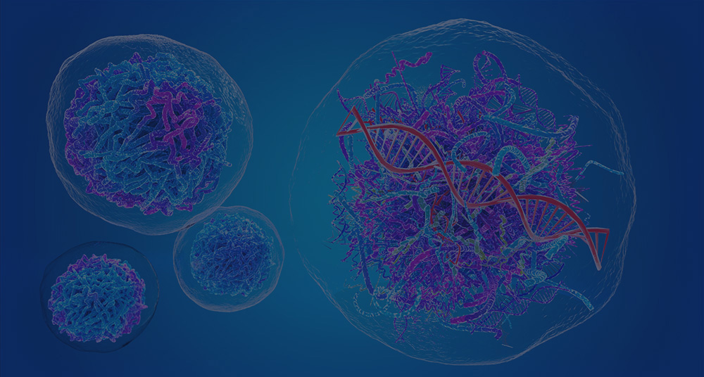
Ribosomal RNA has long been described as the stable scaffold of the translation apparatus. That description is not wrong, but it is no longer enough. In modern molecular biology, rRNA sits at the center of two conversations. The first is mechanistic. rRNA is the structural and catalytic heart of the ribosome. The second is methodological. rRNA is the main reason total RNA libraries fail to capture transcriptome complexity unless enrichment is carefully engineered.
That dual identity matters. If we describe rRNA only as abundant background, we miss its role in ribosome assembly, peptide bond formation, translational selectivity, and chemical modification. If we describe it only as a catalytic scaffold, we miss the practical fact that rRNA can consume most sequencing reads and bury the signal from low-abundance transcripts. Precision RNA-seq depends on handling both truths at once.
This shift is especially important for workflows that cannot rely on poly(A) selection alone. That includes microbial transcriptomics, total RNA profiling, partially degraded inputs, nascent RNA studies, non-coding RNA discovery, and direct RNA approaches. In these settings, rRNA is not a side issue. It is the main determinant of whether sequencing capacity is spent on biology or wasted on molecular bulk. That is why many projects that begin as standard RNA-Seq efforts eventually move toward deeper total RNA sequencing strategies with depletion logic built in from the start.
A useful 2026 article therefore cannot stop at overview language. It must connect ribosome architecture, nucleolar biogenesis, depletion chemistry, and translational regulation into one framework. rRNA is not only abundant. It is organized, catalytic, chemically modified, evolutionarily diverse, and experimentally stubborn.
Ribosomal Architecture: The Catalytic Engine of Life
Why rRNA is more than a scaffold
Proteins often get the credit because they are easier to name and annotate. But in the ribosome, the decisive logic is RNA-centered. The ribosome is a ribonucleoprotein machine, yet its deepest functional core is built around rRNA. That core holds the geometry of decoding, aligns substrates, stabilizes transitional states, and enables catalysis at the peptidyl transferase center.
This changes how we interpret both structure and function. When people describe the ribosome as a protein complex, they often picture proteins forming the active machinery and RNA acting as a passive frame. The opposite is closer to the truth. Ribosomal proteins support folding, assembly, and peripheral stabilization. rRNA defines the catalytic landscape.
The language of “scaffold” is still useful in one narrow sense. rRNA does provide support. It creates the architecture onto which proteins assemble and around which translational factors act. But the word becomes misleading when it suggests passivity. In the ribosome, structure and function are fused. Remove the proteins and support is weakened. Remove the RNA logic and catalysis, decoding geometry, and subunit coordination collapse.
70S versus 80S: divergence without losing the core logic
The most familiar structural split is between the prokaryotic 70S ribosome and the eukaryotic 80S ribosome. The shorthand is useful, but the real difference is not the number. It is the way rRNA and proteins are organized across evolution.
In prokaryotes, the 70S ribosome is formed by the 30S small subunit and the 50S large subunit. In eukaryotes, the 80S ribosome is formed by the 40S small subunit and the 60S large subunit. The naming reflects sedimentation behavior, not simple mass addition. That detail still matters because it reminds us that ribosomes are not stacked parts. Their hydrodynamic behavior emerges from compact three-dimensional organization.
The prokaryotic ribosome tends to look leaner. It carries less peripheral expansion and often presents a stripped-down architecture for readers who want to study core translational logic. The eukaryotic ribosome is larger, more ornamented at the surface, and enriched with additional rRNA expansion segments and associated proteins. Those additions do not erase the ancient catalytic core. They surround it, regulate access to it, and adapt translation to more complex cellular contexts.
Many simplified explanations stop too early. They say prokaryotes have 70S ribosomes and eukaryotes have 80S ribosomes, then move on. But the real biological value lies in interface architecture. The small subunit governs decoding and mRNA-tRNA recognition dynamics. The large subunit contains the peptidyl transferase center and the peptide exit tunnel. The interaction between these modules is not static. It is tuned by conformational motion, factor binding, and the physical layout of rRNA helices and proteins.
The comparison becomes even more useful when designing sequencing experiments across species. In microbial transcriptomics, depletion must accommodate 16S and 23S rRNA targets that may vary across taxa. In eukaryotic total RNA workflows, one must account for 18S, 28S, 5.8S, and 5S species, precursor forms, fragmentation states, and in some cases organellar rRNA carryover. That is why a generic depletion mindset often underperforms when moved from one system to another. Projects that begin with bacterial RNA sequencing needs often expose this problem early, especially when probe designs assume a cleaner rRNA landscape than the sample actually contains.

Figure 1: Comparative architecture of prokaryotic 70S and eukaryotic 80S ribosomes. The conserved lesson is not the sedimentation number but the preservation of an rRNA-centered catalytic core. For experimental design, this matters because depletion targets, precursor forms, and structural accessibility differ across organisms even when the catalytic logic is shared.
The peptidyl transferase center: where RNA performs catalysis
The peptidyl transferase center, or PTC, is the clearest answer to the question: what is rRNA actually doing?
At the PTC, amino acids carried by tRNAs are linked into a growing peptide chain. This is the chemistry that turns genetic information into protein. The active environment of this reaction is shaped by rRNA. The ribosome is therefore a ribozyme in function, even though proteins contribute to assembly and stability elsewhere.
This does not mean the PTC behaves like a free RNA catalyst in solution. The ribosome is an integrated machine. Geometry matters. Local electrostatics matter. The exact positioning of substrates matters. The exit tunnel matters. The timing of tRNA movement matters. rRNA creates a structured catalytic environment in which bond formation becomes both fast and highly constrained.
A useful conceptual move is to stop thinking in binaries. It is not “RNA does everything” or “proteins do everything.” It is that rRNA defines the catalytic logic, while proteins and translational factors shape the operating context. That distinction matters later when we discuss specialized ribosomes and rRNA modification. If the catalytic core is RNA-based, then even subtle variation in rRNA sequence, folding, or chemistry can have functional consequences.
Another important point is that peptide bond formation is not only a chemical event. It is part of a coordinated kinetic cycle. The ribosome must decode the mRNA codon, accept the correct aminoacyl-tRNA, catalyze transfer, translocate, and maintain reading frame. rRNA is involved in this choreography directly or indirectly at multiple steps.
Nucleolar biogenesis: building the molecular machine
If the ribosome is so structurally sophisticated, then its biogenesis must also be highly coordinated. In eukaryotes, that coordination is centered in the nucleolus. Ribosome assembly is not a simple act of transcription followed by spontaneous folding. It is a staged, energy-intensive, highly regulated construction program.
The nucleolus integrates the transcription of precursor rRNA, its processing, its chemical modification, the import of ribosomal proteins, and the stepwise assembly of preribosomal particles. RNA polymerase I produces the large precursor that gives rise to 18S, 5.8S, and 28S rRNAs. RNA polymerase III transcribes 5S rRNA. RNA polymerase II contributes by transcribing mRNAs for ribosomal proteins and assembly factors. These three transcriptional streams must be coordinated in space and time.
This coordination matters for two reasons. First, it explains why ribosome production is tightly linked to growth state, nutrient availability, stress response, and broader cell-state regulation. Second, it shows that rRNA is never just a finished molecule. It exists in precursor, processing, assembled, and modified states. Those states complicate both interpretation and experimental design.
For sequencing specialists, this is not abstract cell biology. It directly affects what total RNA libraries contain. A depletion strategy optimized only for mature cytoplasmic rRNA may fail to remove precursor molecules efficiently. A workflow designed for high-quality cultured RNA may behave differently in stressed samples, tissues with altered nucleolar activity, or degraded and archived research materials. The gap between “known rRNA targets” and “actual sample composition” is one of the most common reasons why depletion underdelivers.
This is also why advanced projects sometimes pair depletion-aware total RNA workflows with long-read or direct-read technologies. When the question is not only abundance but isoform, precursor structure, or chemical state, short-read libraries alone may flatten the picture. In those cases, full-length transcript sequencing by Iso-Seq or nanopore full-length transcript analysis can help restore transcript architecture that fragmentation obscures.
Structural stability is real, but it is not the whole story
rRNA is often described as evolutionarily conserved and structurally stable. That description is true in one narrow sense. The ribosome must preserve core functionality across the tree of life. Large parts of its architecture are therefore highly constrained. But “stable” does not mean “uniform,” and it certainly does not mean “functionally silent.”
There are at least four layers of meaningful variability.
- The first is phylogenetic divergence. Bacterial, archaeal, mitochondrial, chloroplast, and eukaryotic ribosomes all preserve a core logic while differing in sequence, expansion, protein interaction, and assembly pathway.
- The second is processing state. Cells contain mature rRNA, precursor rRNA, partially processed intermediates, and fragments generated by turnover or stress.
- The third is chemical modification. rRNA contains many post-transcriptional marks, including pseudouridylation and 2′-O-methylation, which can influence folding, stability, interaction surfaces, and perhaps translation behavior in context.
- The fourth is population heterogeneity. Even within one cell type, ribosomes may not be perfectly identical. Differences in protein composition, assembly history, modification stoichiometry, or rRNA variant representation may generate ribosome subpopulations with distinct translational biases.
Once we accept that rRNA is both abundant and heterogeneous, we can no longer treat depletion as a crude subtraction step. Depletion becomes a selective physical intervention into a structurally complex RNA population. That is where sequencing strategy begins to matter.
Technical Sovereignty: The Physics and Chemistry of rRNA Depletion
The “90% problem” is really a sensitivity problem
Nearly every total RNA workflow runs into the same obstacle. rRNA dominates the input. In many samples, it represents the overwhelming majority of total RNA mass. That means a sequencing library prepared without proper enrichment logic will spend most of its reads on molecules the investigator did not actually want to quantify.
This is often described as a waste problem, but that wording is too mild. It is a sensitivity problem. When rRNA consumes read space, low-abundance mRNAs become harder to detect. Non-coding RNAs may disappear into sampling noise. Nascent transcripts, partially degraded molecules, and biologically rare species can be lost entirely. The more complex the biological question, the more damaging this compression becomes.
The usual response is to say, “then remove rRNA.” But that answer hides the real challenge. Not all rRNA is equally accessible. Not all samples are equally intact. Not all species share the same rRNA sequence space. Not all workflows want the same remaining RNA population. Good depletion is therefore not a binary step. It is an optimization problem.
This is the point where poly(A) selection and depletion part ways. Poly(A) selection is elegant when the experiment is centered on intact eukaryotic polyadenylated mRNA. It is less useful when the goal includes non-poly(A) transcripts, microbial RNA, degraded RNA, precursor species, or total transcriptome complexity. In those cases, a carefully designed ribodepleted total RNA workflow usually preserves more biology than poly(A)-only enrichment.
In practice, deeper sequencing is not a substitute for better depletion. If the library remains compositionally dominated by rRNA, additional reads mostly reinforce the same imbalance. For low-abundance targets, the more efficient move is often to redesign depletion rather than simply increase depth. For projects that need method support at this stage, custom ribodepletion strategy planning is often more valuable than post hoc rescue.
Why depletion performance varies so much between projects
Many teams are surprised when one depletion workflow works beautifully in one project and only moderately in the next. The reason is that depletion efficiency sits at the intersection of sample chemistry, target sequence, molecular structure, and enzyme behavior.
Start with target abundance. Highly abundant targets are not automatically easy to remove. Abundance helps detection, but it also means small inefficiencies translate into many residual molecules.
Then consider accessibility. rRNA is folded. It forms extensive secondary and tertiary structure and is often complexed with proteins. Probe binding depends not only on complementarity but on whether the target region is physically available. A theoretically perfect probe can perform poorly if its target resides in a structurally protected region.
Fragmentation changes the picture further. In high-quality RNA, long target regions may remain intact and predictable. In degraded RNA, the effective target landscape is broken into shorter pieces. Some probes lose performance because their intended binding sites are truncated. Others gain access because fragmentation opens previously structured regions. This trade-off is easy to underestimate. A probe set optimized for intact molecules may perform differently once the same targets become shorter, more exposed, and less contiguous.
Sequence diversity adds another layer. A depletion probe set designed around reference rRNA may work well for a canonical human sample yet miss organellar contamination, precursor regions, strain-level divergence, or non-model variation. This issue is especially severe in microbiology and metatranscriptomics, where a single sample may contain rRNA pools from many organisms at once. In these settings, depletion often needs to be designed as a population-level solution rather than a single-organism solution, which is why metatranscriptomic sequencing projects frequently benefit from custom rRNA handling logic.
Subtractive hybridization: depletion begins with binding thermodynamics
Subtractive hybridization is often explained in one sentence: probes bind rRNA, then the bound molecules are removed. The sentence is correct but not sufficient. The real engine of this method is hybridization thermodynamics.
A depletion probe must bind strongly enough to outcompete local structure and sample complexity, but not so promiscuously that it captures off-target transcripts. That balance depends on probe length, GC content, mismatch tolerance, salt conditions, temperature, and the folding behavior of both probe and target. In practice, a probe is not simply “complementary” or “not complementary.” It occupies a window of binding probabilities.
This is why probe design should be treated as engineering rather than catalog selection. A short probe may hybridize rapidly but lose specificity. A long probe may improve affinity but become more sensitive to local structure or partial fragmentation. High GC regions can increase stability but also create stubborn self-structure. Low-complexity regions may underperform despite apparent complementarity. Even small sequence mismatches can matter differently depending on where they fall and whether nearby bases stabilize the duplex.
The most successful subtraction strategies therefore think locally. They do not ask only, “Is this rRNA sequence represented?” They ask, “Is this region accessible, robust to fragmentation, tolerant to expected variation, and removable under practical conditions?”
A useful practical framework is to think in terms of three filters. First, is the sequence complementary enough to justify targeting? Second, is the region physically available under real sample conditions? Third, if binding occurs, will the downstream workflow actually convert that event into effective removal? A good depletion design survives all three filters. A weak one often fails at the second or third while looking fine on paper.

Figure 2: rRNA depletion is a selective physical process rather than a generic cleanup step. Probe design must account for abundance, target accessibility, fragmentation state, and duplex stability. For experimental planning, the key failure points are clear: inaccessible targets, unstable hybrids, incomplete cleavage or capture, and library-prep conditions that still favor residual rRNA fragments.
RNase H-mediated cleavage: selective destruction instead of passive removal
RNase H strategies take depletion one step further. Instead of only isolating probe-bound rRNA away from the desired pool, they exploit a biochemical asymmetry. RNase H recognizes RNA-DNA hybrids and degrades the RNA strand within those duplexes. This means the depletion event becomes an active cleavage process.
That small difference has large practical consequences.
- First, cleavage-based depletion can be more flexible across sample classes because it does not rely on one capture geometry alone. Once a stable hybrid forms, the targeted RNA can be selectively destroyed.
- Second, RNase H methods can often be adapted more easily to custom probe panels. This matters when the sample contains unusual rRNA variants, mixed species, precursor material, or noncanonical contaminants that standard kits do not cover well.
- Third, cleavage changes what “success” means. In a capture workflow, success depends on binding and physical separation. In an RNase H workflow, success depends on binding, cleavage efficiency, and the extent to which degraded target fragments no longer dominate the final library. RNase H success is therefore not identical to final-library cleanliness. If cleanup, fragment-size selection, or downstream amplification still enrich residual target pieces, the library can remain noisy even after chemically successful cleavage.
These features make RNase H particularly attractive for species-agnostic or semi-custom applications. They also explain why some advanced service models move beyond one-size-fits-all enrichment and toward project-specific design. A team planning dual RNA-seq, for example, may need to preserve host and microbial informative transcripts simultaneously while suppressing multiple rRNA pools. That is a very different design problem from standard mammalian mRNA profiling.
Poly(A) selection versus rRNA depletion: the real decision framework
The usual comparison table says poly(A) selection enriches mRNA, while rRNA depletion preserves broader transcript diversity. That is true, but it is still too shallow for real project planning. The better question is not which method is superior in general. The better question is which molecular populations must survive library preparation for the biological question to remain answerable.
If the experiment is focused on high-quality eukaryotic mRNA and the goal is coding-transcript quantification, poly(A) selection remains efficient and clean. It reduces library complexity in a useful way. It can lower sequencing burden and simplify interpretation.
But every advantage comes from exclusion. Poly(A) selection discards non-polyadenylated transcripts, weakly polyadenylated species, many non-coding RNAs, and much of the signal from partially degraded or precursor-rich material. It is therefore an optimization for one class of biology, not a neutral default.
rRNA depletion is broader. It keeps more of the transcriptome in play. That makes it well suited for long non-coding RNA analysis, microbial transcriptomics, organellar RNA, precursor RNA, degraded samples, and workflows that need to integrate transcriptional and translational layers. The cost is that library complexity rises and depletion quality becomes a decisive technical variable.
For many modern studies, that trade-off is worth it. If the question touches translational regulation, RNA processing, or noncanonical transcript classes, broader retention is often more valuable than the cleaner simplicity of poly(A) capture. Projects that later expand into polysome sequencing or ribosome-occupancy profiling often benefit from this broader framing, because they are not only asking which transcripts exist. They are asking which transcripts survive, engage, and translate.
Workflow decision guide
| Research objective | Sample property | Keep/remove logic | Preferred workflow |
|---|---|---|---|
| Coding-transcript quantification in intact eukaryotic RNA | High integrity, poly(A)-rich input | Prefer to enrich coding mRNA and reduce complexity | Poly(A) selection |
| Broad transcript discovery including lncRNA and precursor RNA | Mixed RNA classes, non-poly(A) species present | Remove rRNA but retain non-poly(A) molecules | rRNA depletion with total RNA library prep |
| Microbial or mixed-community expression profiling | Diverse species, divergent rRNA targets | Preserve broad transcriptome while targeting multiple rRNA pools | Custom depletion plus metatranscriptomic sequencing |
| Translational regulation or ribosome-state studies | RNA abundance must be compared with ribosome engagement | Retain informative RNA classes and pair with translational readout | rRNA depletion plus matched RNA-seq and ribosome profiling |
Why “dark matter” rRNA breaks standard depletion logic
One of the most underdiscussed technical problems in RNA-seq is what we can call dark matter rRNA. This includes rRNA-derived fragments, precursor species, incompletely annotated variants, organellar contributions, strain-divergent targets, and abundant structured RNA segments that were not fully represented during probe design.
These molecules become visible only when depletion fails. At that point, they are often misread as bad luck, poor sample quality, or random noise. But in many cases they are predictable outcomes of incomplete target modeling.
The fix is not always more sequencing. More sequencing can deepen the same bias. The real fix is better target awareness. That may mean expanding probe coverage, redesigning against structurally accessible regions, stratifying probe sets by species composition, or validating depletion against representative pilot samples instead of assuming reference annotation is enough.
This is where the next generation of direct and modification-aware technologies becomes relevant. If researchers want to know not only what escaped depletion but why it escaped depletion, long-read and direct-read platforms can be informative. Nanopore direct RNA sequencing is especially attractive when the experiment needs to preserve native RNA context, though it introduces its own analytical demands.
Designing depletion around the biological question
One mistake in workflow planning is to treat depletion as a generic preprocessing step with a single success metric. In reality, the right depletion design depends on what the project wants to preserve.
- If the study is trying to recover long non-coding RNAs, then broad transcript preservation matters more than maximal simplification.
- If the study is focused on host-microbe interaction, then the depletion strategy must negotiate multiple rRNA pools without collapsing the dynamic range of informative host and microbial transcripts.
- If the study is trying to capture degraded material, then the design must assume fragmentation from the start and avoid probe logic that only works on long intact targets.
- If the study is moving toward translational analysis, then the depletion strategy must avoid distorting the same RNA classes that later need to be compared with footprint or polysome data.
This is why good RNA-seq planning begins with the biological question rather than the kit shelf. Depletion is not simply a cleanup stage. It is an early design choice that determines which molecular realities remain measurable.
The “Specialized Ribosome” and Epitranscriptomics
Ribosome heterogeneity: from structural conservation to selective translation
For a long time, ribosomes were treated as interchangeable machines. One cell was assumed to contain one functional ribosome class, repeated at scale. That assumption made sense when the main goal was to explain the universal mechanics of translation. It is less useful now. A growing body of work suggests that ribosome populations can vary in composition, assembly history, and chemical state, and that these differences may shape which mRNAs are translated most efficiently under specific conditions.
This is the starting point of the “specialized ribosome” idea. But the phrase is often used too loosely. It should not mean that every ribosome variant creates a fully separate translation program. It should mean something more careful. A specialized ribosome is a ribosome population whose structural or chemical features bias translation toward certain transcript classes, codon contexts, regulatory motifs, or cellular states.
That distinction matters because the field is still separating signal from overinterpretation. Heterogeneity is easier to show than selectivity. It is not hard to detect variation in ribosomal proteins, rRNA modification levels, or assembly intermediates. It is much harder to prove that such variation changes translation in a reproducible and biologically meaningful way.
A rigorous article should not oversell the concept. It should instead describe the evidence ladder. At the lowest level, we see ribosome variation. At the next level, we see ribosome-associated translation bias. At the highest level, we see phenotype: a biased ribosome population shifts protein output in a way that changes cell state, adaptation, or broader phenotypic output.
Where rRNA fits into ribosome specialization
Many discussions of specialized ribosomes focus first on proteins because protein differences are easier to measure. But that framing can miss the deeper layer. rRNA is not only the core scaffold. It is also the chemically modified and structurally tuned substrate on which the ribosome is built.
That means ribosome specialization can emerge from rRNA-linked variables as well as protein-linked variables. These variables include sequence variation across rRNA gene copies or isoforms, differential processing of precursor rRNA, site-specific post-transcriptional modifications, context-dependent modification stoichiometry, and differences in folding stability or subunit maturation.
The most cautious way to phrase this is that rRNA creates a permissive landscape for specialization. It does not need to act alone. A modest shift in rRNA chemistry may alter interaction surfaces, conformational dynamics, decoding behavior, or factor recruitment just enough to change translation preference when combined with the right cellular context.
This is also why the old “structural stability” language needs an update. Structural stability is part of the story. Functional tunability is the other part.
18S and 28S variation: subtle changes, nontrivial consequences
The idea of 18S and 28S rRNA isoforms has become increasingly relevant as researchers look more closely at rDNA copy diversity, tissue-specific expression, and context-dependent ribosome populations. The key insight is that rRNA genes are not always a perfectly homogeneous archive of one sequence. Even when sequence differences are subtle, they can matter if they affect highly connected regions.
A small shift in rRNA sequence does not automatically produce a new translation phenotype. Many sequence differences are probably neutral, buffered, or diluted across the ribosome pool. But neutrality should not be assumed without context.
The effects are most plausible when sequence variation appears near decoding-related regions in the small subunit, functionally important helices, intersubunit interfaces, factor interaction surfaces, or sites that already carry dense chemical modification.
In these regions, even a modest change may alter local folding or interaction geometry. The effect may be too small to notice in bulk translation, yet strong enough to matter for specific transcript subsets, stress states, or developmental transitions.
The right claim is not that every rRNA variant creates a specialized ribosome. The right claim is that rRNA variation expands the possible regulatory space of the translational apparatus.

Figure 3: Ribosome heterogeneity can emerge from isoform diversity, precursor processing, and site-specific modification patterns such as 2′-O-methylation and pseudouridylation. For experimental design, the key bridge is functional readout: heterogeneity matters only when it can be connected to measurable translational bias through footprint positioning, occupancy shifts, or translation-efficiency changes.
rRNA modifications: why Nm and pseudouridylation matter
If sequence variation expands the regulatory space, chemical modification makes that space dynamic.
rRNA is densely modified compared with many other RNA species. Two of the most important and best-studied modification classes are 2′-O-methylation, often abbreviated as Nm, and pseudouridylation, usually written as Ψ. These marks are not decorative. They can influence local flexibility, hydrogen-bonding geometry, folding stability, base stacking, and interactions with proteins or ligands.
That sounds abstract, so it helps to translate the chemistry into functional logic.
A 2′-O-methyl group can stiffen local ribose behavior and alter the physical properties of the RNA backbone. A pseudouridine can change hydrogen-bonding capacity and local structural preferences compared with uridine. In a densely folded RNA such as rRNA, those effects can accumulate in meaningful ways. They can stabilize critical helices, tune interdomain communication, or alter the energetic landscape of the ribosome during translation.
Not every modification site is equally important. Some likely maintain structural integrity with little condition-specific variability. Others may vary in occupancy or context, making them stronger candidates for regulatory behavior. This is why the modern question is no longer just “Where are the modifications?” It is “Which sites vary, under what conditions, and with what translational consequences?”
This question has pushed more groups toward dedicated modification-aware assays, including 2′-O-RNA methylation sequencing for Nm-sensitive projects and broader nanopore RNA methylation sequencing approaches when native RNA context is important.
Mapping 2′-O-methylation: what RiboMeth-seq really contributes
RiboMeth-seq has become one of the defining methods for profiling Nm in rRNA. Its value lies in how it converts chemical stability into a measurable signal. Sites carrying 2′-O-methylation show altered cleavage behavior under controlled conditions, allowing computational inference of methylation patterns across the RNA.
The appeal of the method is clear. It can deliver site-level maps at scale and it aligns naturally with the abundance of rRNA. But its strengths are often described too vaguely. The real contribution of RiboMeth-seq is not that it merely “finds methylation.” It provides a structured way to compare relative protection profiles across samples, which can be used to infer site occupancy and detect differential methylation patterns.
Still, no method is complete on its own. RiboMeth-seq depends on experimental consistency, cleavage behavior, coverage, and threshold setting. Sites with partial occupancy can be especially challenging. So can contexts where structural features or sample quality distort the expected readout. Mapping is not the same as direct truth capture. It is model-based inference built on a chemical signal.
Mapping pseudouridylation: why Ψ remains methodologically tricky
Pseudouridylation is functionally important and analytically stubborn. Unlike canonical uridine, pseudouridine is an isomer. It carries the same mass as uridine, but its glycosidic linkage is rearranged. That makes direct detection harder than many readers expect.
Historically, many Ψ-mapping methods have relied on selective chemical derivatization or reverse transcription behavior. These strategies can identify candidate positions, but they often require careful controls and conservative interpretation. Signal strength may depend on sequence context, local structure, reaction efficiency, and data-processing thresholds.
This is why pseudouridylation mapping often remains more interpretive than introductory articles admit. A site may be detectable in one assay and weak in another. A change in signal may reflect biology, but it may also reflect coverage, chemistry, or analysis stringency.
A useful technical article should therefore avoid simplistic wording such as “this method directly measures all Ψ sites.” A better formulation is that different assays provide different windows into pseudouridylation, with different balances of throughput, site specificity, and confidence.
Native nanopore sequencing: the promise and the caution
Native nanopore sequencing has changed the modification conversation because it offers something other methods often do not: the possibility of reading RNA molecules in a near-native state, one molecule at a time, without mandatory cDNA conversion.
That matters for rRNA because modifications do not exist in isolation. They exist on structured molecules with specific sequence contexts and processing states. In principle, nanopore data can preserve more of that context. It can help distinguish mature from precursor molecules, support long-range interpretation across the same RNA molecule, and open the door to modification-aware single-molecule analysis.
That promise is real, but the caution is just as important.
Nanopore-based modification calling is not a simple lookup exercise. Signal deviations must be interpreted through models that are sensitive to local k-mer context, training data quality, basecalling assumptions, and algorithm choice. Different pipelines may disagree. Some predicted sites are robust. Others are model-dependent. This is especially important for readers who assume that direct RNA sequencing automatically means unambiguous modification calling.
The right conclusion is not to lower confidence in nanopore methods. It is to use them correctly. Nanopore sequencing is strongest when treated as a context-rich measurement platform that benefits from orthogonal validation, careful controls, and transparent thresholding. For studies that need native RNA context together with long-range transcript structure, researchers often combine direct RNA nanopore workflows with full-length long-read transcript support.
Modification maps are not endpoints. They are mechanistic hypotheses.
One of the biggest analytical mistakes in this area is to treat a modification map as the finish line. It is not. A map is the start of mechanistic questioning.
Once Nm or Ψ sites are identified, the real work begins. Are these sites constitutive or variable? Do they change with stress, differentiation, or experimental perturbation? Are they enriched in functional hotspots? Do they alter ribosome assembly or mature ribosome behavior? Do they correlate with selective translation of defined transcript classes?
Only when those questions are asked does epitranscriptomic mapping become translational biology rather than site cataloging.
This is also the point where multi-layer study design becomes valuable. Bulk RNA abundance alone is not enough. Researchers increasingly need combinations such as total RNA-seq, direct RNA analysis, structural inference, and translational readouts. That broader logic fits naturally with multi-omics study design when a single assay cannot resolve mechanism on its own.
Ribosome profiling: from occupancy to translational control
Ribosome profiling, or Ribo-seq, remains one of the most powerful ways to connect ribosome behavior to transcript output. The basic idea is elegant. Ribosome-protected RNA fragments are isolated and sequenced, allowing researchers to map where active ribosomes sit on transcripts.
The first value of Ribo-seq is positional. It can reveal which transcripts are being translated and where ribosomes accumulate.
The second value is kinetic. It can expose initiation bottlenecks, elongation pauses, codon-sensitive behavior, upstream open reading frame usage, and context-dependent changes in ribosome density.
The third value is comparative. When paired with matched RNA-seq, it allows estimation of translation efficiency, often abbreviated as TE. In simple terms, TE compares ribosome occupancy with transcript abundance. A transcript with high RNA abundance but modest ribosome engagement has a different regulatory profile from a transcript with modest abundance but very strong ribosome loading.
This makes Ribo-seq central to the specialized ribosome discussion. If rRNA heterogeneity or modification state truly shifts translational preference, some part of that effect should become visible in ribosome-protected footprint patterns, TE values, or context-specific ribosome distribution.
That does not mean Ribo-seq directly proves specialized ribosomes by itself. It does not. But it provides a genome-wide readout of translational consequence, which is exactly what modification maps and ribosome heterogeneity studies need. For matched assay planning, paired RNA-seq and translational footprinting workflows are often the most informative starting point.
Translation efficiency: powerful metric, easy to misuse
Translation efficiency is useful, but it should be handled with care. The common shorthand is:
TE = ribosome footprint abundance / RNA abundance
That expression is helpful, but it is not a full mechanistic truth. TE is a composite metric. It reflects multiple processes at once, including initiation rate, ribosome loading, elongation behavior, transcript stability, footprint recovery bias, and library normalization choices.
This means TE should not be interpreted as a direct proxy for “how strongly a gene is translated” without context. A high TE value may reflect efficient initiation. It may also reflect slower elongation and footprint accumulation. A low TE value may reflect weak translation. It may also reflect rapid ribosome transit or changes in RNA stability.
The solution is not to avoid TE. The solution is to interpret it alongside positional footprint data, transcript class, experimental design, and biological context. In practice, the strongest translational studies use TE as one layer rather than the only layer. Replicate structure also matters. A striking TE shift without stable replicate behavior or footprint-position support should be treated cautiously.
Integrating depletion, modification mapping, and translational readout
At a strategic level, these topics should no longer be planned in isolation. Depletion determines which RNA molecules survive into the dataset. Modification mapping determines how rRNA chemical states are represented. Translational readouts determine whether those states matter functionally. If any one layer is designed poorly, the others become harder to interpret.
Consider a study on stress-induced translational rewiring. If depletion removes informative precursor or noncoding species unevenly, RNA abundance baselines become distorted. If modification calling is noisy, candidate regulatory sites become ambiguous. If Ribo-seq lacks matched RNA context, translational shifts lose interpretive grounding. The result is a fragmented story. The solution is coordinated experimental logic.
Researchers who begin with broad transcript retention often later need matched RNA-seq plus translational readout planning rather than isolated assays, especially when the end goal is to connect RNA-state heterogeneity to functional protein-output bias.
A practical framework for precision RNA-seq in rRNA-heavy systems
At this point, the central message becomes clear. rRNA should be handled as three things at the same time.
- First, it is the catalytic and structural core of the ribosome. Without that recognition, ribosome biology becomes oversimplified.
- Second, it is a heterogeneous molecular substrate. Sequence variation, precursor state, and chemical modification create functional diversity that may influence translation.
- Third, it is the dominant technical barrier in total RNA sequencing. Without well-designed depletion, many of the most interesting RNA species remain under-sampled.
These three truths can be turned into a practical planning framework.
If the project is centered on coding mRNA abundance in clean eukaryotic RNA, poly(A) selection may still be sufficient.
If the project involves degraded samples, non-coding RNAs, microbial systems, precursor transcripts, or translational regulation, rRNA depletion should be considered early rather than added as a rescue step.
If the project asks mechanistic questions about ribosome state, then depletion strategy, modification mapping, and translational readout should be designed as a connected system, not as independent modules.
A useful final test is simple: which RNA molecules must remain visible for the biological claim to survive? If the answer includes non-polyadenylated RNAs, precursor states, structured rare species, or translationally regulated targets, then broad retention and smart depletion become essential. If the answer includes ribosome-state biology itself, then rRNA is not just background. It is part of the signal.
FAQ
What is the main biological role of rRNA in the ribosome?
rRNA forms the structural and catalytic core of the ribosome. It helps define the geometry of decoding, supports subunit architecture, and shapes the peptidyl transferase center where peptide bond formation occurs.
Why is rRNA depletion so important in RNA-seq?
Because rRNA often dominates total RNA input. Without depletion, a large fraction of sequencing reads is spent on highly abundant ribosomal molecules, reducing sensitivity for mRNA, non-coding RNA, precursor RNA, and other low-abundance species.
When is rRNA depletion better than poly(A) selection?
rRNA depletion is usually the better choice when the study needs to retain non-polyadenylated RNAs, degraded RNA, microbial RNA, precursor transcripts, or broad total transcriptome complexity. Poly(A) selection is better suited to intact eukaryotic mRNA-focused studies.
What is meant by a specialized ribosome?
A specialized ribosome is a ribosome population whose composition or chemical state biases translation toward certain transcript classes or regulatory contexts. The term should be used carefully and ideally supported by evidence for translational selectivity, not just structural variation.
Why are 2′-O-methylation and pseudouridylation important in rRNA?
These modifications can influence local RNA structure, stability, and interaction behavior. In a large and highly folded RNA such as rRNA, those effects may alter ribosome assembly, functional tuning, or context-dependent translation.
What does RiboMeth-seq measure?
RiboMeth-seq infers 2′-O-methylation patterns by using cleavage behavior and sequencing-based readout to identify sites that show methylation-associated protection signatures.
Can nanopore sequencing directly detect rRNA modifications?
Nanopore sequencing can provide modification-sensitive signal patterns on native RNA molecules, but interpretation depends on computational models, training quality, and validation strategy. It is powerful, but it should not be treated as automatically unambiguous.
What is translation efficiency in Ribo-seq analysis?
Translation efficiency generally compares ribosome footprint abundance with RNA abundance for the same transcript. It is useful for identifying shifts in translational control, but it must be interpreted carefully because it reflects several overlapping biological and technical factors.
How should researchers choose between RNase H depletion and bead-based capture?
The key decision is not brand preference but failure mode. RNase H is often attractive when custom probe design, mixed-species input, or cleavage-based selectivity is needed. Bead-based capture can work well when target binding and physical separation are sufficient. In both cases, final-library composition matters more than nominal depletion chemistry.
What is the best workflow logic for degraded or mixed-species RNA inputs?
Start by defining which RNA classes must remain measurable. Then design depletion around fragmentation state, target diversity, and downstream assay needs. For mixed-species or heavily fragmented inputs, broad-retention workflows with custom depletion logic are usually more reliable than generic defaults.
References
- Ramakrishnan V. “The Ribosome Emerges from a Black Box.” Cell. 2014;159(5):979-984. 10.1016/j.cell.2014.10.052
- Natchiar SK, Myasnikov AG, Hazemann I, Klaholz BP. “Visualization of chemical modifications in the human 80S ribosome structure.” Nature. 2017;551:472-477. 10.1038/nature24482
- Lafontaine DLJ, Riback JA, Bascetin R, Brangwynne CP. “The nucleolus as a multiphase liquid condensate.” Nature Reviews Molecular Cell Biology. 2021;22:165-182. 10.1038/s41580-020-0272-6
- Parker MD, Doyle M, et al. “Efficient and specific oligo-based depletion of rRNA.” Scientific Reports. 2019;9:12281. 10.1038/s41598-019-48692-2
- Orelle C, Carlson ED, Szal T, Florin T, Jewett MC, Mankin AS. “Protein synthesis by ribosomes with tethered subunits.” Nature. 2015;524:119-124. 10.1038/nature14862
- Genuth NR, Barna M. “The discovery of ribosome heterogeneity and its implications for gene regulation and organismal life.” Molecular Cell. 2018;71(3):364-374. 10.1016/j.molcel.2018.07.018
- Norris K, Hopes T, Aspden JL. “Ribosome heterogeneity and specialization in development.” WIREs RNA. 2021;12(4):e1644. 10.1002/wrna.1644
- Shi Z, Fujii K, Kovary KM, Genuth NR, Rost HL, Teruel MN, Barna M. “Heterogeneous ribosomes preferentially translate distinct subpools of mRNAs genome-wide.” Molecular Cell. 2017;67(1):71-83.e7. 10.1016/j.molcel.2017.05.021
- Erales J, Marchand V, Panthu B, et al. “Evidence for rRNA 2′-O-methylation plasticity: control of intrinsic translational capabilities of human ribosomes.” PNAS. 2017;114(49):12934-12939. 10.1073/pnas.1707674114
- Krogh N, Jansson MD, Häfner SJ, et al. “Profiling of 2′-O-Me in human rRNA reveals a subset of fractionally modified positions.” Nature Communications. 2016;7:11483. 10.1038/ncomms11483
- Schwartz S, Motorin Y. “Next-generation sequencing technologies for detection of modified nucleotides in RNAs.” RNA Biology. 2017;14(9):1124-1137. 10.1080/15476286.2016.1251543
- Begik O, Lucas MC, Liu H, et al. “Quantitative profiling of pseudouridylation dynamics in native RNAs with nanopore sequencing.” Nature Biotechnology. 2021;39:1278-1291. 10.1038/s41587-021-00915-6
- Jenjaroenpun P, Wongsurawat T, Wadley TD, et al. “Decoding the epitranscriptional landscape from native RNA sequences.” Nucleic Acids Research. 2021;49(2):e7. 10.1093/nar/gkaa620
- Ingolia NT. “Ribosome footprint profiling of translation throughout the genome.” Cell. 2016;165(1):22-33. 10.1016/j.cell.2016.05.044
- McGlincy NJ, Ingolia NT. “Transcriptome-wide measurement of translation by ribosome profiling.” Methods. 2017;126:112-129. 10.1016/j.ymeth.2017.05.028
- Choi J, Ieong KW, Demirci H, et al. “N6-methyladenosine in mRNA disrupts tRNA selection and translation-elongation dynamics.” Nature Structural & Molecular Biology. 2016;23:110-115. 10.1038/nsmb.3291
- Ferretti MB, Ghalei H, Ward EA, Potts EL, Karbstein K. “Rps26 directs mRNA-specific translation by recognition of Kozak sequence elements.” Nature Structural & Molecular Biology. 2017;24:700-707. 10.1038/nsmb.3447
- Leppek K, Das R, Barna M. “Functional 5′ UTR mRNA structures in eukaryotic translation regulation and how to find them.” Nature Reviews Molecular Cell Biology. 2018;19:158-174. 10.1038/nrm.2017.103
Related Services
For research use only. Not for use in diagnostic procedures.