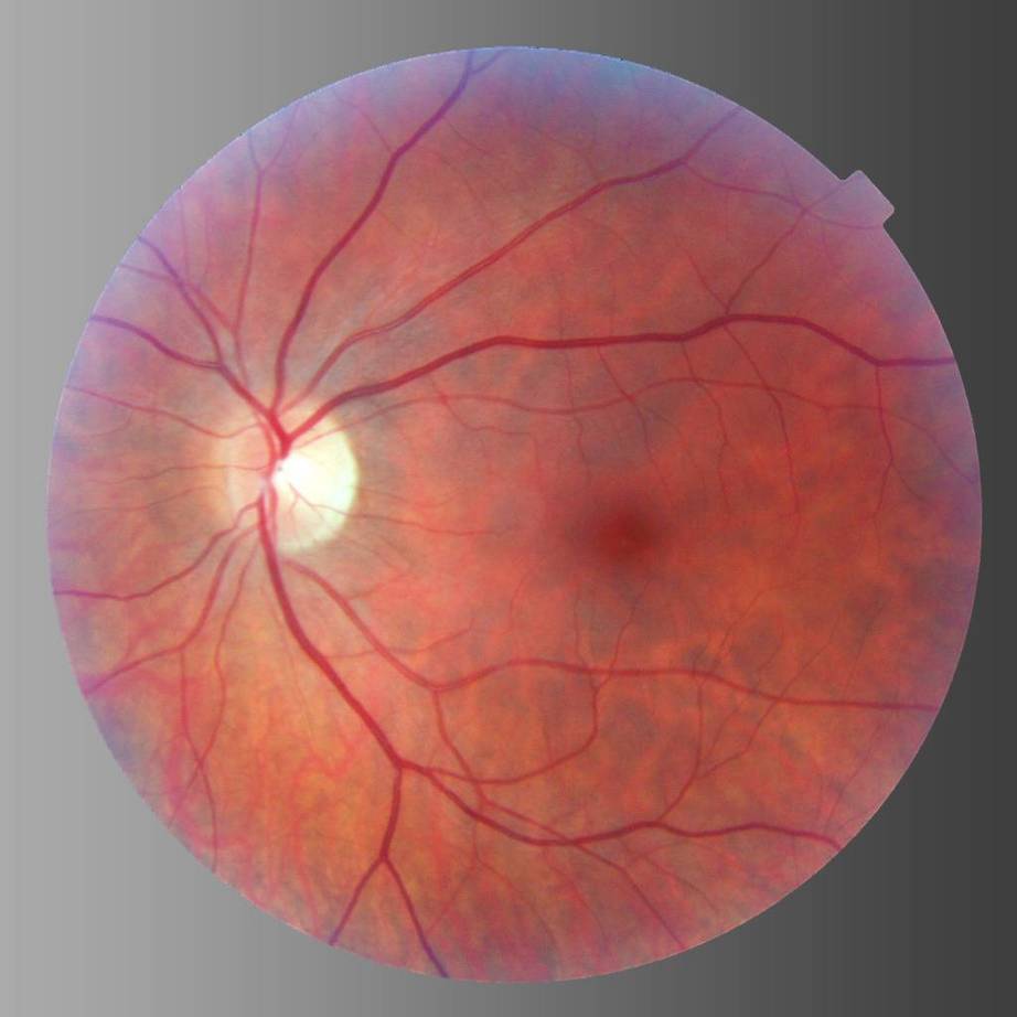
Vitreoretinopathy is an autosomal and X-linked genetic eye disorder with pathological features associated with vitreous and retinal degeneration, which can cause irreversible loss of vision. Early phenotypes of the disease include decreased vision, cataracts, retinal detachment, glaucoma, neovascularization and peripheral fibro-vascular proliferation, and traction retinal detachment. These familial diseases include Wagner syndrome, Stickler syndrome, Knobloch syndrome, Norrie disease, retinoschisis and exudative vitreoretinopathy. Vitreoretinopathy has high variability in clinical phenotypes, involving mutations of approximate 250 genes. And this disease can be induced by secondary complications such as diabetes. However, there is no effective treatment for this disease.
Vitreoretinopathy is an ocular disorder caused by genetic mutations. Many decisive genes involved in this disease are located in chromosome 11. Bestrophin-1 encoded by BEST1 gene is highly expressed in the retina, which is involved in the membrane integrity. The mutation in BEST1 gene has been verified to be associated with more than five types of retinopathy. The protein encoded by CAPN5 gene is a calcium-dependent protein kinase involved in signal transduction. The mutations of CAPN5 gene alter its enzymatic activity that causes neovascular inflammatory. ZNF408 is associated to exudative vitreoretinopathy and retinal pigmentation if homozygous mutations occur. And the protein regulated by FZD4 is involved in the development of retinal blood vessels in vertebrates. Mutations of FZD4 fail to response Norrin induced signaling and further lead to Familial exudative vitreoretinopathy (FEVR). Structural protein family coding gene COL is related to Stickler Syndrome, Knobloch Syndrome and Marshall Syndrome. Other genes such as KIF11, KCNJ13, LRP5, NR2E3, NDP, RS1, TSPAN12, VCAN, etc. are also related to vitreoretinopathy.
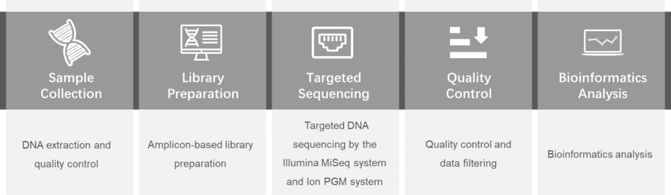
Our platform provides the comprehensive and efficient targeted DNA Sequencing by the Illumina MiSeq system/Ion PGM system to verify the potential pathogenic nucleotide variants. We also offer detailed vitreoretinopathy-related gene list, from which you can choose genes you are interested in for genetic testing.
| BEST1 | CAPN5 | ZNF408 | FZD4 |
| ATOH7 | COL11A2 | COL18A1 | COL2A1 |
| COL9A1 | COL9A2 | COL9A3 | CTC1 |
| CTTNNB1 | KCNJ13 | KIF11 | NDP |
| NR2E3 | P3H2 | RS1 | TSPAN12 |
| VCAN | LRP5 | RS1 |
For more information about the Custom Vitreoretinopathy Panel or need other amplification requirements, please contact us.
References:
Please submit a detailed description of your project. We will provide you with a customized project plan to meet your research requests. You can also send emails directly to for inquiries.
Please fill out the form below: ×