Systemic lupus erythematosus (SLE) is an autoimmune disease that invades the connective tissue of the body. The lesions often involve multiple systems and organs. It is characterized by the production of large amounts of autoantibodies and immune complex deposition. While SLE can occur in both males and females, it is found far more often in women, and the symptoms associated with each sex are different. The pathogenesis of SLE is complex. Genetic factors and environmental factors are involved in the pathogenesis of SLE. At present, it is considered that the imbalance of apoptotic nuclear fragmentation and clearance in SLE leads to the exposure of various nuclear antigens to the immune system, and the adaptive immune response is activated by antigen presenting cells (APC), thereby breaking the immune balance. In addition, APC can release a variety of cytokines to promote the development of autoimmune reactions, thereby aggravating the disease.
Since the occurrence of SLE involves in multiple genetic regions, it may have oligomeric properties, meaning that it is possible for several genes to jointly regulate the susceptibility of the disease. BANK1 gene encodes a specific scaffold protein for B cells. Its activated form can affect intracellular calcium mobilization induced by B cell receptors. This may contribute to sustained B-cell receptor signaling and make B cells overactive, causing systemic lupus erythematosus. In addition, researchers have identified a genetic association between signal transduction and transcriptional activator 4 (STAT4) in SLE. Induced by IL-12 produced by dendritic cells, STAT4 is an essential regulator of the immune system. It leads to the development of Th1 cells which have the capacity to secrete high levels of IFN-γ. In addition, STAT4 also transmits a variety of key cytokine-inducing signals, including interleukin-12 and interleukin-23. Therefore, mutations in STAT4 may largely affect the immune response and cause autoimmunity in humans. Furthermore, IRF5, PTPN22, CDKN1A, ITGAM, BLK are TNFSF4 are also associated with the occurrence of SLE.
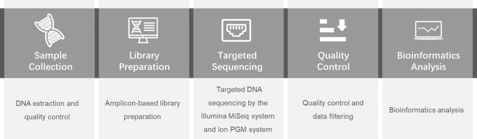
Based on the latest research reports, we select a variety of genes that are highly associated with increased risk of systemic lupus erythematosus. You can select the genes you want to detect in the panel library or customize your exclusive panel. We offer specialized targeted sequencing technologies to detect low frequency changes in genes that may be mutated.
| BANK1 | BCR | BLK | CCL2 | CCL19 |
| CD4 | CD25 | CR2 | CTLA4 | C4A |
| C4B | C4B-2 | CXCL10 | FCGR2B | GTF2I |
| HCY | HDL | IL-1β | IL-18 | IRF5 |
| IRAK4 | LAG3 | LTK | MIF | NCF2 |
| ORF2p | P40 | P450 | PDCD1 | PXK |
| P2RX7 | PTPN22 | RASGRP1 | RTK | RIPK1 |
| STAT4 | TAM | TLR5 | TNFSF4 | TNFAIP3 |
| TREX1 | TLR7 |
For more information about the Custom Systemic Lupus Erythematosus Panel or need other amplification requirements, please contact us.
References:
Please submit a detailed description of your project. We will provide you with a customized project plan to meet your research requests. You can also send emails directly to for inquiries.
Please fill out the form below: ×