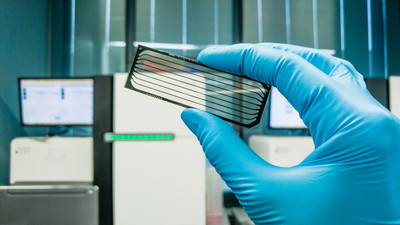
Core Steps in the eCLIP-seq Protocol: A Detailed Guide to Mapping RNA-Protein Interactions
The dynamic interaction between RNA binding protein (RBP) and RNA is the core driving force of a series of life activities in cells, and its function runs through the whole process of gene expression regulation—from RNA processing after transcription initiation (such as splicing, capping and tailing) to intracellular transportation and localization of RNA, to the start and end of translation and degradation and metabolism of RNA, and every link is inseparable from the precise regulation of RBP.
Enhanced Cross-Linking and Immunoprecisions followed by Sequencing (eCLIP-seq) technology, as an innovative tool to analyze this kind of interaction, has achieved a multi-dimensional upgrade compared with the traditional CLIP technology: its single nucleotide resolution can be accurate to the binding event of a single base on RNA, High specificity eliminated more than 90% of non-specific binding signals through size matching input control and strict washing steps, while standardized experimental procedures improved the reproducibility of results between different laboratories to more than 80%, so it quickly became the mainstream research method in this field.
This article details the core steps of the eCLIP-seq protocol, including cell lysis, UV crosslinking, immunoprecipitation, RNA adapter ligation, library preparation, sequencing, and data analysis, for mapping RNA-protein interactions.
Introduction to eCLIP-seq Workflow
The core purpose of eCLIP-seq is to map the interaction between RBP and RNA with high resolution. In life activities, the interaction between RBP and RNA is extremely complex and precise, and it is often difficult for traditional techniques to accurately capture the details of this interaction. eCLIP-seq can locate the binding site of RBP and RNA in the whole genome through a series of optimized experimental steps and data analysis methods:
- Cell Lysis and UV Crosslinking: Lyse cells using a mild buffer containing detergents and protease inhibitors to preserve complex integrity. Irradiate cells with 254nm ultraviolet light to covalently crosslink RNA-binding proteins (RBPs) with their bound RNA, stabilizing the complexes.
- Immunoprecipitation: Use specific antibodies against target RBPs, coupled to magnetic beads, to enrich RBP-RNA complexes. Implement stringent washing steps to reduce non-specific background. Parallelly process size-matched input (SMI) controls to correct technical biases.
- Adapter Ligation: Sequentially ligate barcoded 3' and 5' adapters to RNA fragments. Unique barcodes enable sample multiplexing and error correction in subsequent analyses.
- Reverse Transcription and PCR Amplification: Convert RNA to cDNA using reverse transcriptase, accounting for potential termination at crosslink sites. Amplify cDNA with a limited number of PCR cycles to avoid over-amplification artifacts.
- High-Throughput Sequencing: Perform paired-end sequencing with a recommended depth of 20–50 million reads to ensure sufficient coverage of binding sites.
- Bioinformatics Analysis: Process raw data to identify RBP binding sites, and motif analysis.
 SHlFTR delivers a high-resolution view on the protein interactions of differentsequence regions in the SARS-CoV-2 RNA genome (Aydin et al., 2024)
Service you may intersted in
Learn More:
Cell Lysis and UV Crosslinking for eCLIP
In the eCLIP-seq experimental process, cell lysis and UV crosslinking are the key steps to lay the experimental foundation, and the accuracy of their operation directly affects the quality of subsequent separation and analysis of RNA-protein complexes.
Crosslinking of RNA-protein Complex
UV irradiation (254nm) is the core means to realize a covalent connection between RBPs and binding RNA. When cells are irradiated by 254nm ultraviolet rays, the pyrimidine bases in RNA molecules will undergo a photochemical reaction with amino acid residues in protein to form covalent bonds, thus stably crosslinking RBPs with their bound RNA. This cross-linking method is highly specific and can only act on RNA and protein that directly interact with each other, which ensures that the complex studied in the follow-up experiment is a real physiological interaction product and avoids the interference of non-specific binding on the experimental results.
Cell Lysis and RNase Treatment
The cell lysis adopts a mild lysis method, which aims to break the cell membrane and release the cell contents while preserving the integrity of the RNA-protein complex to the greatest extent. Mild lysis buffer usually contains a proper amount of detergent (such as NP-40) and a protease inhibitor, which can not only effectively destroy the cell structure, but also inhibit the activity of protease and prevent protein degradation, thus maintaining the stable state of the complex.
After cell lysis, controlled RNase digestion is needed to achieve RNA fragmentation. RNase specifically cleaves RNA molecules and breaks them down into fragments suitable for subsequent experimental analysis. By controlling the concentration of RNase, reaction time, and temperature, RNA fragments with moderate length can be obtained, which can not only carry enough binding site information but also facilitate the subsequent linker connection and sequencing process.
Key Considerations
Optimizing the crosslinking time is a crucial link in this step. If the crosslinking time is too short, the covalent connection between RNA and protein is not sufficient, which will lead to the loss of some real interaction complexes in the subsequent treatment, which will affect the sensitivity of the experiment. However, if the cross-linking time is too long, it may lead to excessive fragmentation of RNA and the loss of effective information.
At the same time, it may also damage the structure of protein and RNA and interfere with subsequent immunoprecipitation and sequencing analysis. Therefore, in the experimental process, it is necessary to determine the best cross-linking time through pre-experiments, so as to balance the cross-linking efficiency and the integrity of the complex and provide high-quality samples for the subsequent experimental steps.
 Development of a scalable, unbiased and highly efficient method to identify proteins bound to specific RNA regions in endogenously expressed RNAs (Aydin et al., 2024)
Immunoprecipitation of RBP-RNA Complexes
Immunoprecipitation (IP) is the core step to isolate and enrich the target RBP-RNA complex in the eCLIP-seq experiment, and its efficiency and specificity directly determine the quality of subsequent experimental data. This process mainly includes key links such as antibody selection and magnetic bead coupling, controlling washing stringency, and setting size matching input (SMI) control.
- A. Antibody selection and magnetic bead coupling
- a) Selecting highly specific antibodies (verified by IP) is the basis of successful immunoprecipitation. Such antibodies can accurately recognize and bind to target RBP, and minimize the binding to non-target proteins, thus improving the specificity of complex separation. At the same time, coupling antibodies with magnetic beads is an important means to achieve efficient separation.
- b) Magnetic microspheres have a large specific surface area, can be stably combined with antibodies, and can be quickly separated under the action of a magnetic field, which is convenient for subsequent washing and complex recovery operations. Through the coupling of antibody and magnetic beads, the antibody-magnetic bead complex can specifically capture the RBP-RNA complex in the sample, which lays the foundation for the subsequent purification steps.
- c) A strict washing step is the key to reducing the nonspecific background, which is also an important improvement of eCLIP-seq compared with the traditional CLIP technology. In the process of immunoprecipitation, in addition to the target RBP-RNA complex, there are a lot of nonspecific binding proteins, RNA, and other impurities in the sample. These nonspecific binding substances can be effectively removed by using washing buffers with different concentrations, such as salt solutions and detergents, many times.
- B. SMI comparison
- a) Parallel processing of input samples to control technical deviation is a highlight of eCLIP-seq experimental design, that is, SMI control. The input sample refers to the part of the cell lysate that has not undergone the immunoprecipitation step, which contains all RNA and protein components in the sample. By performing the same treatment on the input sample as the immunoprecipitation sample, such as RNA fragmentation and linker ligation, and comparing it with the data of the immunoprecipitation sample, the technical deviation caused by factors such as RNA fragment size difference and sequencing preference in the experimental process can be effectively corrected. The setting of SMI control improves the accuracy and reliability of the experimental results and makes the identification of RBP binding sites more accurate.
 CLIP variants for studying RBP-RNA interactions (Lin et al., 2019)
RNA Adapter Ligation and Library Preparation
RNA linker ligation and library preparation are the key links in the eCLIP-seq experiment to transform captured RNA fragments into a testable sequence library, which directly affects the quality of sequencing data and the accuracy of subsequent analysis.
3' and 5' Joint Connection
In this step, the unique bar-coded connectors are connected to the 3' and 5' ends of RNA fragments by sequential connection. The unique barcode connector can not only distinguish different samples and avoid cross-contamination between samples, but also help identify and correct errors in subsequent data analysis. The sequential connection ensures the effective combination of the linker and both ends of the RNA fragment, provides necessary primer binding sites for subsequent reverse transcription and PCR amplification, and also helps to maintain the integrity and directionality of the RNA fragment, laying a foundation for accurately analyzing the binding sites of RNA and protein.
Reverse Transcription Challenge
In the process of reverse transcription, the main challenge is to deal with the phenomenon of reverse transcription termination caused by crosslinking, which is also a feature of eCLIP sequencing reading. Because UV cross-linking makes RNA and protein form covalent bonds, these cross-linking sites may hinder the progress of reverse transcriptase when it moves along the RNA template, leading to the early termination of the reverse transcription process and the production of shorter cDNA fragments. These reverse transcription termination sites caused by cross-linking are just important markers of RNA binding to protein, but they also bring difficulties to the smooth progress of the reverse transcription reaction.
Researchers need to optimize reverse transcription reaction conditions, such as adjusting enzyme concentration, reaction temperature, and time, to minimize the impact of these termination phenomena on cDNA synthesis, and at the same time, make full use of this feature to locate binding sites in data analysis.
PCR Amplification and Size Selection
PCR amplification adopts a limited number of cycles to avoid the deviation caused by over-amplification. The limited number of cycles can reduce the distortion of fragment proportion caused by the difference in amplification efficiency while ensuring the acquisition of sufficient DNA. After amplification, gel extraction was carried out to enrich the optimal fragment size of 100-300 nt.
Fragments of this size range not only contain enough information about RNA binding sites, but also meet the sequencing requirements of a high-throughput sequencing platform. Size selection by gel extraction can remove too long or too short fragments and non-specific amplification products, improve the purity and uniformity of the library, and ensure that the sequencing data accurately reflects the interaction between RNA and protein.
 A comparison of small RNA library preparation workflows (Shore et al., 2016)
Sequencing and Data Analysis of eCLIP
Sequencing and data analysis are the key links to transform the original experimental materials into biologically meaningful information in the eCLIP-seq experiment, and their rationality and rigor directly determine the reliability and scientificity of the research results.
High-throughput Sequencing
In high-throughput sequencing, the recommended sequencing depth is about 200-50 million reads. This depth range can ensure that enough information about RNA binding sites is covered, and at the same time, it can avoid the waste of resources caused by too deep sequencing. At the same time, using double-ended sequencing technology, that is, sequencing the two ends of DNA fragments, can provide more sequence information than single-ended sequencing, which is helpful to more accurate sequence comparison and subsequent data analysis, and improves the accuracy of RNA binding site positioning.
Bioinformatics Process
The bioinformatics process includes several key steps, which together complete the transformation from original sequencing data to biological conclusions.
- First, the linker pruning is performed using tools such as Cutadapt to remove the linker sequence contained in the sequencing data. These linker sequences were introduced in the process of library preparation, and they do not belong to the RNA sequence of the sample itself. If they are not removed, they will interfere with the subsequent analysis.
- Followed by alignment, using tools such as STAR and Bowtie2 to align the trimmed sequence to the reference genome or transcriptome. By comparison, the position of RNA fragments obtained by sequencing in the genome or transcriptome is determined, which provides a basis for finding binding sites in the future.
- Then, the peak recognition, with the help of CLIPper, PyPeaks, and other tools, was used to identify binding sites. By analyzing the distribution of sequencing readings on the genome, these tools find out the areas with rich readings, that is, the possible binding sites of RNA binding proteins, which are called "peaks".
- Finally, motif analysis, using tools such as HOMER and MEME, is used to study the specificity of RNA-binding proteins. By analyzing the nucleotide sequence around the binding site, we can find out the characteristic short sequences (motifs), which are usually the key sequences for RNA-binding proteins to recognize and bind to RNA, and can reflect the binding preference of the protein.
 NGS data comparison between CleanTag and TruSeq Small RNA Library PreparationKit (Shore et al., 2016)
Conclusion
eCLIP-seq workflow fixes RNA-protein interaction through cell lysis and UV crosslinking, separates specific complexes through immunoprecipitation, and realizes efficient and accurate identification of RNA-protein binding sites through RNA linker connection, library preparation, sequencing, and data analysis. The optimization of this process makes the experimental results more reliable, which provides strong technical support for an in-depth study of the function of RNA-binding proteins and the molecular mechanism of RNA-protein interaction, and promotes the research progress in related fields.
References
- Aydin J, Gabel A, Zielinski S, et al. SHIFTR enables the unbiased identification of proteins bound to specific RNA regions in live cells. Nucleic Acids Res. 2024 52(5): e26.
- Lin C, Miles WO. Beyond CLIP: advances and opportunities to measure RBP-RNA and RNA-RNA interactions. Nucleic Acids Res. 2019 47(11): 5490-5501.
- Shore S, Henderson JM, Lebedev A, et al. Small RNA Library Preparation Method for Next-Generation Sequencing Using Chemical Modifications to Prevent Adapter Dimer Formation. PLoS One. 2016 11(11): e0167009.