BACS-Seq: A Comprehensive Guide to Cyclization-Based Pseudouridine Detection
BACS-Seq (2-bromoacrylamide-assisted cyclization sequencing) is a base-resolution method for detecting pseudouridine (Ψ), the most abundant RNA modification in cells. Developed by the Song group at the University of Oxford and published in Nature Methods in 2024, BACS-Seq uses a cyclization reaction that converts Ψ sites into U-to-C mutations during reverse transcription — a fundamentally different signal from the truncations and deletions produced by older methods. This article explains how the chemistry works, where BACS-Seq outperforms CMC-based and bisulfite-based approaches, what the method has already revealed about the human epitranscriptome, and how to choose the right Ψ detection strategy for different research questions.
Key Takeaways:
- BACS-Seq produces a U-to-C mutation signal at Ψ sites, enabling true single-base resolution even in consecutive uridine tracts and densely modified regions where bisulfite-based methods (BID-seq, PRAISE) lose positional accuracy.
- The method achieves quantitative accuracy that correlates strongly with mass spectrometry (r = 0.90 vs SILNAS), compared to r = 0.37 for BID-seq and r = 0.48 for PRAISE.
- In its landmark 2024 study, BACS-Seq detected all 105 known Ψ sites in human rRNA, mapped 609 high-confidence Ψ sites across human cytosolic tRNAs, and revealed 304 Ψ sites in human snoRNAs — far exceeding the coverage of prior methods.
- The technology was developed by Chun-Xiao Song's laboratory at Oxford and is described in the peer-reviewed literature; the underlying chemistry is the subject of patent applications held by the Ludwig Institute for Cancer Research.
- For researchers evaluating Ψ detection strategies, the choice among BACS-Seq, BID-seq, PA-Ψ-seq, CMC-based methods, and nanopore direct RNA sequencing depends on RNA type, required resolution, quantitative needs, and sample availability.
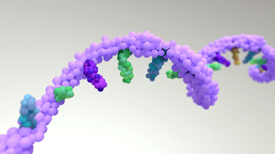
 Figure 1. The core BACS-Seq chemistry — 2-bromoacrylamide selectively reacts with pseudouridine (Ψ) to form a cyclized adduct that produces a U-to-C mutation signature during reverse transcription.
What BACS-Seq Measures
Pseudouridine (Ψ) is not a standard base call in any sequencing pipeline, yet it is the single most abundant modified nucleotide in cellular RNA. Often called the fifth nucleotide, Ψ appears in tRNA, rRNA, snRNA, snoRNA, and mRNA, where it strengthens RNA secondary structure, stabilizes codon-anticodon interactions, and — in the case of mRNA — can promote stop codon readthrough with direct implications for therapeutic mRNA design.
Pseudouridine as the Fifth Nucleotide
Unlike methylation marks that add chemical groups to existing bases, Ψ is an isomer of uridine: the uracil base is rotated and reattached at the C5 position rather than N1. This single atomic rearrangement changes everything about how the base behaves — it forms an extra hydrogen bond, rigidifies the sugar-phosphate backbone, and alters the way reverse transcriptase reads the modified site. These properties make Ψ biologically important and analytically frustrating. The mass of Ψ is identical to uridine (244.2 Da for the nucleoside), so standard mass spectrometry cannot distinguish them without chromatographic separation. And because Ψ still base-pairs with adenine in most contexts, standard RNA-seq sees nothing at all.
Why Base Resolution Matters
The difference between knowing that a tRNA contains "several Ψ modifications somewhere in the TΨC loop" and knowing exactly which positions carry Ψ — and at what stoichiometry — is the difference between correlative observation and mechanistic insight. Pseudouridine synthases (PUS enzymes) act on specific substrate positions. In cancer, mutations in PUS enzymes including DKC1 (dyskerin) drive disease through altered rRNA pseudouridylation. In mRNA therapeutics, Ψ substitution at specific positions alters immunogenicity and translation efficiency. Each of these questions demands base-resolution, quantitative Ψ detection. For a broader view of how RNA modification detection technologies have evolved, see the RNA modification detection technologies overview.
The Cyclization Chemistry
The core innovation of BACS-Seq is a chemical reaction that exploits the one structural feature unique to Ψ among all canonical and modified nucleosides: a free N1 position. In uridine, N1 carries the glycosidic bond to ribose. In Ψ, that bond moves to C5, leaving N1 exposed and nucleophilic.
How 2-Bromoacrylamide Targets Ψ
The BACS reaction uses 2-bromoacrylamide as a bifunctional reagent. The acrylamide moiety acts as a Michael acceptor: the free N1 of Ψ attacks the terminal alkene, forming a covalent N1-acrylamide adduct. The α-bromine then serves as a leaving group for an intramolecular O2-alkylation, creating a stable five-membered cyclized product termed nce1,2Ψ (carbamido-1,O2-ethano-Ψ). Unmodified uridine, which lacks the free N1, cannot initiate the first Michael addition and remains unreacted. The reaction runs in phosphate buffer at 85°C for 30 minutes — conditions mild enough to preserve RNA integrity while driving the cyclization to near-completion. After two rounds of purification to remove unreacted reagent, the RNA is ready for reverse transcription.
| Reaction Component | Role in BACS Chemistry |
|---|---|
| 2-Bromoacrylamide | Bifunctional reagent: Michael acceptor (acrylamide) + leaving group (α-bromine) |
| Ψ free N1 | Nucleophile; initiates Michael addition — absent in uridine |
| Phosphate buffer (pH ~7) | Maintains neutral pH for selective Ψ reaction |
| 85°C, 30 min | Reaction temperature and duration for >85% conversion |
| nce1,2Ψ product | Stable cyclized adduct; read as cytosine during RT |
From Chemical Adduct to U-to-C Mutation
The cyclized nce1,2Ψ product alters the Watson-Crick face of the base. During reverse transcription, the modified base is no longer recognized as uridine. Instead, the reverse transcriptase inserts a dGTP opposite the adduct, producing a guanosine in cDNA where the original RNA carried Ψ. When this cDNA is sequenced and aligned to the reference genome, the Ψ position appears as a T-to-C mismatch (thymine in the reference, cytosine in the read). The mutation rate at each candidate site directly reflects Ψ stoichiometry: a site with 80% modification produces an ~80% C call rate, while an unmodified uridine shows only background-level mutation. Across 256 tested sequence motifs, the average Ψ-to-C conversion rate was 87.6%, and the average false-positive rate on unmodified uridine was 0.75%.
 Figure 2. Three detection paradigms for pseudouridine — CMC-based methods produce RT truncations with positional ambiguity; bisulfite-based methods produce deletion signals that cannot resolve consecutive Ψ sites; BACS-Seq produces a point mutation that uniquely identifies each modified position.
Where Older Ψ Methods Struggle
Before BACS-Seq, transcriptome-wide Ψ detection fell into two chemical categories, each with a fundamental limitation that the field worked around for over a decade.
CMC-Based Methods and the Truncation Problem
N-cyclohexyl-N′-(2-morpholinoethyl)carbodiimide (CMC) reacts with Ψ at the N3 position under alkaline conditions. The CMC-Ψ adduct blocks reverse transcriptase, producing truncated cDNA at or near the modification site. Methods including Pseudo-seq, Ψ-seq, and CeU-seq all rely on this principle. The problems are well documented: CMC labeling efficiency is low and variable across sequence contexts; the truncation signal is inherently imprecise — reverse transcriptase may stall one or two nucleotides before or after the actual Ψ, creating a "stutter" pattern that blurs positional assignment; and CMC also reacts, albeit more slowly, with uridine and guanosine, producing background that must be filtered statistically across 14 or more biological replicates. The result is a qualitative method — it can report that Ψ is present somewhere in a transcript, but it cannot reliably report the modification fraction at a specific nucleotide.
Bisulfite-Based Methods and the Deletion Blind Spot
Bisulfite-induced deletion sequencing (BID-seq), published in Nature Biotechnology in 2023, improved on CMC methods by enabling quantitative Ψ detection from as little as 10–20 ng of poly(A)+ RNA. Under neutral pH, bisulfite selectively reacts with Ψ to form a Ψ-bisulfite adduct without deaminating cytidine — preserving sequence complexity. During reverse transcription, the adduct causes the enzyme to skip the modified site, producing a deletion in the cDNA. The deletion rate correlates with Ψ stoichiometry.
The limitation is geometric. When two or more Ψ modifications sit in consecutive uridines — a common arrangement in rRNA expansion segments and tRNA loop regions — the deletion signal cannot determine which uridine was modified. The alignment algorithm assigns the deletion to the 5′-most uridine by default, but this is a computational convention, not a measurement. In practice, bisulfite-based methods (BID-seq and PRAISE) lose single-base resolution in the very regions where Ψ is most abundant. This limitation is structural — it cannot be fixed by deeper sequencing or improved calibration curves.
BACS-Seq solves this problem by producing a point mutation rather than a deletion. Each U-to-C transition is assigned to a specific nucleotide position during alignment, regardless of whether adjacent uridines are also modified. In the 28S rRNA expansion segment containing Ψ3737, Ψ3741, Ψ3743, Ψ3747, and Ψ3749 — five modifications within a 13-nucleotide window — BACS-Seq resolved all five independently, while bisulfite-based methods could not distinguish individual sites.
BACS Against Other Detection Methods
The following table summarizes the four main approaches to transcriptome-wide Ψ detection, their signal types, and their key performance characteristics.
| Feature | CMC-Based (Pseudo-seq, Ψ-seq) | BID-seq | PRAISE | BACS-Seq | Nanopore DRS |
|---|---|---|---|---|---|
| Signal type | RT truncation | Deletion | Deletion | U-to-C mutation | Ionic current shift |
| Base resolution in poly-U | No (stutter) | No (deletion ambiguity) | No (ambiguous peaks) | Yes | Partial |
| Quantitative (stoichiometry) | No | Partial (r=0.37 vs MS) | Partial (r=0.48 vs MS) | Yes (r=0.90 vs MS) | Yes (per-read) |
| Dense Ψ region detection | Poor | Poor | Poor | Good | Moderate |
| Simultaneous A-to-I detection | No | Partial | No | Yes | Yes |
| RNA input | ≥2 μg poly(A)+ | 10–20 ng poly(A)+ | ~100 ng | ~100 ng–1 μg | ~50–500 ng |
| Chemical treatment | CMC (harsh) | Bisulfite (harsh) | Bisulfite (harsh) | Bromoacrylamide (mild, 85°C) | None |
Quantitative Accuracy Compared to Mass Spectrometry
The gold standard for Ψ quantification is SILNAS (stable isotope-labeled NA SILAC mass spectrometry), which measures global Ψ stoichiometry but provides no positional information. Comparing each sequencing method's reported Ψ levels against SILNAS measurements in human rRNA reveals the quantitative accuracy gap. BACS-Seq achieves a Pearson correlation of r = 0.90 with SILNAS data across all 105 known rRNA Ψ sites. BID-seq achieves r = 0.37; PRAISE achieves r = 0.48. The difference matters: at r = 0.37, a site with 60% true Ψ stoichiometry might be reported anywhere from 10% to 90%, making the quantitative claim unreliable for biological interpretation.
Resolving Consecutive Uridine and Dense Ψ Regions
The structural advantage of a point-mutation signal over a deletion signal becomes clearest in the most heavily modified RNAs. Human U2 snRNA contains 14 Ψ sites, several in close proximity. BACS-Seq resolved all 14 — the first high-throughput sequencing method to do so. In human 28S rRNA, five consecutive modifications within the Ψ3737–Ψ3749 cluster were all individually resolved. In human mitochondrial tRNAs, BACS-Seq detected 65 Ψ sites, compared to 34 for PRAISE (of which only 13 were resolved with both quantitation and single-base resolution). These are not marginal improvements — they represent entirely new biochemical information that was previously inaccessible.
 Figure 3. The BACS-Seq bioinformatics analysis pipeline from raw sequencing reads through Ψ site calling, annotation, and differential modification analysis.
Turning Mutations into Modification Maps
Once the cyclization reaction and sequencing are complete, the bioinformatics task is conceptually straightforward but requires careful thresholding: identify every position in the transcriptome where the U-to-C mutation rate exceeds the sequence-context-specific background, then quantify the modification fraction from the excess mutation rate.
Ψ Site Calling and Annotation
The analysis pipeline begins with standard RNA-seq preprocessing — adapter trimming, quality filtering, and alignment to the reference genome and transcriptome. After alignment, the pipeline scans every reference thymine position (corresponding to uridine in the original RNA) and tallies the base calls from aligned reads. A position is flagged as a candidate Ψ site when the C-call fraction exceeds a background threshold determined from unmodified control libraries or from the same sequence motif in transcripts where Ψ is absent. Because the false-positive rate varies slightly across the 256 possible NNΨNN sequence motifs, the filtering step applies motif-specific background models. Candidate sites are then annotated by RNA biotype — tRNA, rRNA, snoRNA, snRNA, mRNA, or other non-coding RNA — using reference transcript databases.
For researchers working with tRNA modification data, the annotation step must account for the extensive nucleotide modifications that tRNAs carry beyond Ψ, which can affect both alignment and mutation calling. Dedicated tRNA analysis workflows use modified-reference-aware alignment strategies.
Differential Modification Analysis
Once Ψ sites are identified and quantified across samples, the analysis follows a differential modification framework analogous to differential expression analysis in RNA-seq. For each Ψ site, the modification fraction is compared between conditions using statistical models that account for read depth, biological variability, and the sequence-context-specific mutation background. The output is a list of differentially modified Ψ sites, each with a fold-change and statistical confidence metric.
Downstream interpretation typically includes motif enrichment analysis around differentially modified sites, integration with RNA expression data to distinguish modification changes from abundance changes, and functional enrichment of the transcripts carrying altered Ψ levels. For researchers requiring pseudouridine detection through alternative chemistry, photoactivatable approaches offer complementary signal types that can serve as orthogonal validation.
Discoveries Powered by BACS
The 2024 Nature Methods paper by Xu, Kong, Cheng, and colleagues demonstrated BACS-Seq's capabilities across a comprehensive set of RNA biotypes.
The First Quantitative Human tRNA Ψ Map
Cytosolic tRNAs are the most heavily pseudouridylated RNA species, but prior methods had failed to produce a reliable quantitative Ψ map. CMC-based methods performed poorly on highly structured tRNAs, and bisulfite-based methods could not resolve closely spaced Ψ sites in tRNA loop regions. BACS-Seq identified 609 high-confidence Ψ sites across human cytosolic tRNAs, with modification hotspots at positions 13, 27–28, 38–40, and 55 — all functionally critical regions of the tRNA tertiary structure. Position 55, in the TΨC loop that gives the loop its name, was the most frequently modified and most heavily modified site across the entire tRNA repertoire. In mitochondrial tRNAs, BACS-Seq detected 65 Ψ sites, nearly double the 34 reported by PRAISE.
Complete snoRNA and snRNA Ψ Landscapes
The improvement in coverage was even more dramatic in small nucleolar and small nuclear RNAs. BACS-Seq detected 304 Ψ sites in HeLa snoRNAs, compared to 11 sites reported by Ψ-seq and 39 by BID-seq — an order-of-magnitude increase. In the spliceosomal snRNAs, BACS-Seq resolved all 14 Ψ sites in U2 snRNA, something no prior high-throughput method had achieved. Two consecutive Ψ sites at positions 11 and 12 in U4atac snRNA were individually resolved, demonstrating the method's ability to handle the most spatially challenging modification arrangements.
mRNA Pseudouridine and Stop Codon Readthrough
In HeLa poly(A)+ RNA, BACS-Seq identified 1,335 Ψ sites across the mRNA transcriptome. More than half (55.3%) of these sites resided in consecutive uridine sequences — precisely the sequence context where bisulfite-based methods cannot assign exact positions. Ψ sites in mRNA showed enrichment in coding sequences and 3′ untranslated regions, with a sequence motif preference for GUΨCN and poly-U contexts.
A finding with direct relevance to mRNA therapeutics: Ψ was detected within stop codons of mammalian transcripts. This observation supports a mechanistic model in which pseudouridylation of stop codon uridines promotes translational readthrough — a phenomenon exploited in mRNA vaccine design, where Ψ substitution enhances protein expression. Understanding exactly which positions carry Ψ in endogenous transcripts provides a reference map for optimizing therapeutic mRNA sequences.
The following table summarizes the key quantitative achievements of BACS-Seq across RNA biotypes in the landmark 2024 study.
| RNA Biotype | Ψ Sites Detected (BACS-Seq) | Prior Best Method (sites) | Key Finding |
|---|---|---|---|
| Cytosolic rRNA (5.8S, 18S, 28S) | 103 of 105 known + 4 novel | SILNAS MS (105 known, no positional data) | r = 0.90 correlation with MS quantification |
| Cytosolic tRNA | 609 high-confidence | No prior quantitative map existed | Hotspot positions: 13, 27–28, 38–40, 55 |
| Mitochondrial tRNA | 65 | PRAISE: 34 (only 13 with true base resolution) | ~2× improvement in site detection |
| snoRNA | 304 | BID-seq: 39; Ψ-seq: 11 | ~8–28× improvement in coverage |
| U2 snRNA | 14 (all known sites) | No prior method resolved all 14 | First complete U2 Ψ map by HTS |
| HeLa mRNA | 1,335 | BID-seq: 575 | 55.3% of sites in consecutive-U contexts |
Picking the Right Ψ Detection Strategy
There is no single best method for every pseudouridine research question. The right choice depends on what RNA you are studying, what resolution you need, how much sample you have, and whether you need quantitative stoichiometry or simply presence/absence calls.
| Research Goal | Recommended Approach | Why |
|---|---|---|
| Initial screening for Ψ presence across the transcriptome | BID-seq or CMC-based method | Lower cost; sufficient for presence/absence; BID-seq adds quantitation |
| Base-resolution Ψ mapping in tRNA or rRNA | BACS-Seq | Only BACS-Seq resolves consecutive-U and dense Ψ regions in highly structured RNAs |
| Quantitative Ψ stoichiometry for mechanism studies | BACS-Seq | r = 0.90 correlation with gold-standard MS; only method with verified quantitative accuracy |
| Ultra-low-input samples (pg–ng range) | Uli-epic BID-seq | Optimized BID-seq variant works with as little as 100 pg mRNA |
| Simultaneous detection of Ψ + other modifications | Nanopore direct RNA sequencing | Single-molecule, multi-modification information without chemical treatment |
| Detection of Ψ + A-to-I editing + m1A in one experiment | BACS-Seq | BACS-Seq simultaneously captures all three modification types |
| Orthogonal validation of Ψ sites | PA-Ψ-seq or targeted qPCR | Independent chemical principle for confirmation |
| Service-based Ψ detection without in-house method development | BID-seq or PA-Ψ-seq | Available as established RNA modification services with standardized workflows |
FAQ
How does BACS-Seq compare to BID-seq for mRNA Ψ detection?
Both methods provide transcriptome-wide, base-resolution Ψ detection with quantitative information. BACS-Seq offers higher quantitative accuracy (r = 0.90 vs r = 0.37 correlation with mass spectrometry) and unambiguous resolution of Ψ sites in consecutive uridine tracts — where over 55% of mRNA Ψ sites are located. BID-seq requires less input RNA (10–20 ng vs ~100 ng–1 μg) and benefits from a longer track record of published applications across diverse biological systems. For projects where every mRNA Ψ site must be precisely mapped regardless of sequence context, BACS-Seq provides more complete information. For projects with limited RNA input or where the research question does not require resolving closely spaced Ψ sites, BID-seq offers a well-validated alternative.
Can BACS-Seq data be analyzed with standard RNA-seq bioinformatics tools?
Not directly. BACS-Seq requires a specialized analysis pipeline that identifies U-to-C mutations above sequence-context-specific background thresholds. Standard RNA-seq aligners and variant callers are not optimized for the chemically induced, modification-dependent mutation signature that BACS-Seq produces. The published method includes custom analysis scripts for Ψ site calling, annotation, and differential modification testing. Researchers working with Ψ detection data — regardless of which sequencing method generated it — should ensure their bioinformatics pipeline accounts for the specific error models and background characteristics of the chosen chemistry.
References
- Xu H, Kong L, Cheng J, et al. "Absolute quantitative and base-resolution sequencing reveals comprehensive landscape of pseudouridine across the human transcriptome." Nature Methods, vol. 21, no. 11, 2024, pp. 2024–2033. doi:10.1038/s41592-024-02439-8
- Dai Q, Zhang LS, Sun HL, et al. "Quantitative sequencing using BID-seq uncovers abundant pseudouridines in mammalian mRNA at base resolution." Nature Biotechnology, vol. 41, no. 3, 2023, pp. 344–354. doi:10.1038/s41587-022-01505-w
- Zhang LS, Ye C, Ju CW, et al. "BID-seq for transcriptome-wide quantitative sequencing of mRNA pseudouridine at base resolution." Nature Protocols, vol. 19, no. 2, 2024, pp. 517–538. doi:10.1038/s41596-023-00917-5
- Luo N, Huang Q, Zhang M, Yi C. "Functions and therapeutic applications of pseudouridylation." Nature Reviews Molecular Cell Biology, vol. 26, no. 9, 2025, pp. 691–705. doi:10.1038/s41580-025-00852-1
- Ron K, Kahn J, Malka-Tunitsky N, Sas-Chen A. "High-throughput detection of RNA modifications at single base resolution." FEBS Letters, vol. 599, no. 1, 2025, pp. 19–32. doi:10.1002/1873-3468.15052
- Ding H, Liu N, Wang Y, et al. "Implications of RNA pseudouridylation for cancer biology and therapeutics: a narrative review." Journal of Translational Medicine, vol. 22, no. 1, 2024, 906. doi:10.1186/s12967-024-05687-6
- He W, Xu C, Chen W, et al. "Uli-epic: profiling RNA modifications from ultra-low input samples." Genome Biology, vol. 26, 2025, 384. doi:10.1186/s13059-025-03857-3
- Xiong J, Wu J, Liu Y, Feng YJ, Yuan BF. "Quantification and mapping of RNA modifications." Trends in Analytical Chemistry, vol. 172, 2024, 117606. doi:10.1016/j.trac.2024.117606
Research Use Only. The information provided in this article is for research and educational purposes only. CD Genomics provides research-use-only epigenomics and epitranscriptomics sequencing services.