3′UTR CpG Islands in Cancer: Beyond Promoter-Centered DNA Methylation
Most discussions of CpG islands and cancer DNA methylation begin at gene promoters. In a 2026 Life Science Alliance study, Wilson and Kanhere examined a less visible region: CpG islands located in 3' untranslated regions, where methylation may relate to gene expression, transcript-end regulation, and cancer-specific regulatory patterns. The article was published online on July 24, 2026, as Life Science Alliance 9(10):e202503234 (doi:10.26508/lsa.202503234; PMID:42498499).
This article reviews the published study’s main observations, including the shorter and less CpG-dense but conserved nature of 3'UTR CGIs and their enrichment for factors such as ZFP57. It places those findings beside earlier work on 3'UTR methylation and alternative polyadenylation, then outlines the evidence needed to test whether a 3'UTR methylation signal is a useful regulatory lead rather than a correlation produced by the surrounding tumor epigenome.
Key Takeaways
- 3'UTR CpG islands represent a small but distinct genomic population that should not be merged automatically with promoter CGIs or the broader gene body.
- The study linked 3'UTR CGI methylation patterns with cancer-associated expression changes, but the direction and strength of the association varied by cancer type and locus.
- CTCF-dependent alternative polyadenylation provides a plausible mechanistic bridge, yet 3'UTR methylation-expression associations still require locus-specific and perturbational validation.
- A useful follow-up project combines region-aware methylation analysis with RNA-seq, isoform or poly(A) measurements, and targeted chromatin or methylation assays.
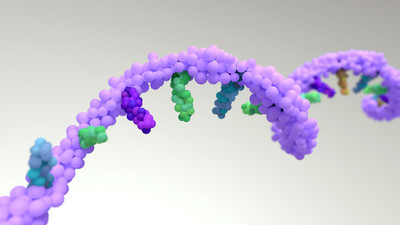
 Figure 1: A promoter-centered view of DNA methylation can miss regulatory information near the 3' end of genes.
The Missing 3'UTR Layer
The familiar CpG-island model is promoter-centered. A promoter CGI is often discussed as a regulatory switch: when it remains unmethylated, transcription-factor access and transcriptional activity are compatible; when it becomes aberrantly methylated, gene silencing can follow. That model remains useful, but it does not describe every CGI in the genome.
3'UTRs sit downstream of the coding sequence and are usually discussed as RNA regulatory regions. Their transcripts can contain microRNA and RNA-binding-protein sites that affect stability, localization, and translation. The corresponding genomic region also lies near transcription termination and polyadenylation decisions. Treating the DNA sequence in this region as biologically irrelevant simply because it is not a promoter leaves out a possible layer of regulation.
This distinction matters in cancer datasets. A gene may show an expression change even when its promoter methylation is stable. If methylation probes in the 3'UTR are pooled into a broad “gene body” category, a locus-specific pattern can disappear inside a much larger annotation class. Region-aware annotation is therefore a first analytical decision, not a cosmetic relabeling step.
For projects that need genome-wide coverage across promoter, gene-body, and 3'UTR regions, researchers can review the genome-wide DNA methylation analysis service as a starting point for study design.
A Distinct CGI Population
Wilson and Kanhere identified 909 CpG islands, approximately 3% of all annotated CGIs, that overlapped 3'UTRs without a nearby promoter CGI. These sites were associated with hundreds of protein-coding genes. The exact count depends on the genome build, annotation priority, transcript models, and exclusion rules, so the number should be treated as an analysis-defined population rather than a universal constant.
The more consequential observation was the contrast between structural compactness and evolutionary conservation. The 3'UTR CGIs were generally shorter and had lower CpG density than promoter CGIs, yet their conservation was comparable to or higher than the promoter CGI comparison set. This combination makes them easy to overlook in a simple CGI census while still suggesting that the sequence context may be under functional constraint.
The study also reported a different baseline methylation state. Promoter CGIs are often protected from methylation in normal cells, whereas the 3'UTR CGI group showed high methylation in both normal and tumor samples. That does not mean methylation is automatically functional at every site. It means that “CGI equals unmethylated promoter-like element” is an unsafe assumption when the CGI is located at the 3' end of a gene.
| Feature | Promoter CGI | 3'UTR CGI in the study | Interpretation |
|---|---|---|---|
| Typical genomic context | Near a transcription start site | Within or overlapping a 3'UTR | Region annotation changes the biological question |
| CpG density and length | Often higher and longer | Generally shorter and less CpG-dense | A lower-density CGI can still be conserved |
| Baseline methylation pattern | Often protected from methylation | Frequently methylated in normal and tumor samples | Methylation status cannot be inferred from CGI label alone |
| Main regulatory question | Transcription initiation | Expression association, transcript-end processing, or chromatin effects | Mechanism needs locus-specific testing |
The site’s Human & Mouse CpG Island Panel Sequencing page is relevant when a project begins with CGI-focused discovery. The panel should not be presented as automatically resolving every 3'UTR question; transcript annotation and the exact capture design still determine which loci are measured.
 Figure 2: Promoter and 3'UTR CGIs can differ in location, CpG density, baseline methylation, and the regulatory questions they support.
Cancer Patterns Are Contextual
The cancer analysis focused on breast cancer and hepatocellular carcinoma, where matched normal and tumor methylation data were available. Across both groups, 3'UTR CGI probes were highly methylated overall, while promoter CGI probes remained comparatively unmethylated. The meaningful changes appeared at subsets of CpG sites rather than as a uniform shift across every 3'UTR CGI.
The direction of change was not the same in both cancer types. Breast cancer showed a stronger tendency toward hypermethylation at the highlighted 3'UTR CGI sites, whereas hepatocellular carcinoma showed both hypermethylated and hypomethylated changes. This is a useful warning against treating “3'UTR hypermethylation” as a pan-cancer rule. The relevant unit may be a cancer type, a cell state, a locus, or a subset of CpGs within the CGI.
The study then connected differentially methylated 3'UTR regions with gene-expression changes. Earlier work supports the broader premise that 3'UTR methylation should be analyzed separately: McGuire and colleagues reported enrichment of positive methylation-expression associations in 3'UTRs across tumor datasets and linked one example, Havcr2, to T-cell activation. These findings support a regulatory hypothesis, but they also underline the need to consider cell composition and transcriptional state when using bulk tumor data.
For an analysis that combines differential methylation with gene expression, the Epigenomic Data Analysis service is a natural internal resource. When the question includes transcript abundance or isoform behavior, Integrating RNA-seq and Epigenomic Data Analysis is the more specific route to review.
The LRFN1 Example
LRFN1 illustrates why the promoter-centered model is not always enough. In the study, breast and liver tumor data contained a differentially methylated region within the 3'UTR CGI of LRFN1, while the promoter CGI did not show the same change. The 3'UTR methylation pattern was associated with increased LRFN1 expression in the analyzed tumor settings.
This is a useful research case because the methylation and expression signals occupy different regulatory layers. It does not establish that the 3'UTR methylation change causes the expression increase. Tumor purity, cell-state composition, copy-number effects, transcriptional feedback, and altered RNA processing could all contribute to the observed relationship.
The practical value of the case is therefore not a finished mechanism. It is a prioritization rule: when a gene’s promoter methylation does not explain its expression change, inspect the 3'UTR and other non-promoter regions before concluding that methylation is unrelated to the phenotype.
For a short list of candidate loci, a targeted DNA methylation analysis service can be considered for focused confirmation after discovery-level analysis. The target list should be defined from independent evidence and matched transcript annotations rather than selected only because a single CpG has a large tumor-normal difference.
Mechanisms Still Need Testing
The most useful mechanistic connection comes from work on DNA methylation and alternative polyadenylation. Nanavaty and colleagues showed that demethylation at specific genomic regions can permit CTCF binding, recruit cohesin, and promote chromatin looping that changes proximal versus distal polyadenylation-site usage. The study used molecular perturbations and cellular assays, so it provides a mechanism for selected loci rather than a universal explanation for all 3'UTR CGIs.
That distinction is important. A 3'UTR methylation signal could relate to transcript-end processing, RNA polymerase II elongation, binding of methylation-sensitive factors, chromatin organization, or a downstream consequence of high transcription. The same methylation-expression direction can therefore arise through different routes in different loci.
Wilson and Kanhere also highlighted ZFP57 as a candidate factor based on motif and binding-data analyses. This is a strong hypothesis-generating result, not a completed causal chain. A mechanistic claim would need perturbation of the candidate factor or methylation state, a measurable change in chromatin or transcript-end behavior, and a locus-specific rescue or orthogonal validation.
 Figure 3: A mechanism-oriented follow-up connects regional methylation signals to chromatin, transcript-end, and perturbation evidence.
| Evidence layer | What it can support | What it cannot establish alone |
|---|---|---|
| Differential 3'UTR methylation | A cancer-associated regional signal | That methylation initiates the expression change |
| Methylation-expression correlation | A relationship worth prioritizing | Direction of causality or molecular mechanism |
| ChIP-seq or CUT&Tag for CTCF, ZFP57, or related factors | Factor occupancy near the candidate region | That occupancy is methylation-dependent without perturbation |
| RNA-seq with isoform or poly(A) analysis | Changes in expression and transcript-end usage | That the methylation signal is the direct driver |
| Targeted methylation and perturbation assays | Locus-specific evidence for a regulatory model | Generalization to all 3'UTR CGIs |
Researchers considering factor-occupancy evidence can review ChIP-Seq, CUT&Tag, and the site’s epigenomic transcription factor binding site analysis resources as complementary options. The appropriate assay depends on the factor, sample type, expected occupancy, and whether the project needs discovery-scale or locus-focused evidence.
What a Follow-Up Study Needs
A follow-up study should be designed around the claim it needs to support. If the aim is to find 3'UTR methylation candidates, genome-wide profiling and careful annotation may be enough for the discovery stage. If the aim is to explain a cancer-associated expression change, the project needs matched molecular layers and a validation path that separates correlation from mechanism.
| Study question | Useful evidence | Main interpretation risk |
|---|---|---|
| Which 3'UTR CGIs differ between tumor and matched normal tissue? | Region-aware methylation profiling, matched samples, DMR analysis | Cell composition or batch effects can mimic locus-specific change |
| Does methylation track gene expression? | Methylation plus RNA-seq from the same samples | Correlation can reflect transcriptional state or tumor purity |
| Does the signal alter transcript-end processing? | Poly(A)-site or isoform analysis, 3' end RNA sequencing, transcript models | Incomplete annotation can misclassify alternative UTRs |
| Does a candidate factor bind the region? | ChIP-seq, CUT&Tag, motif analysis, public binding tracks | Binding does not prove methylation dependence |
| Is the locus causal? | Targeted methylation manipulation, factor perturbation, rescue or reporter assay | A single-locus result may not generalize across cancers |
Three design choices are especially important.
- Keep the annotation explicit. Record genome build, CGI definition, transcript source, and the rule used to prioritize promoter, 5'UTR, exon, intron, and 3'UTR overlaps.
- Match the molecular layers. When possible, use methylation and RNA data from the same samples or clearly matched groups. Add isoform or poly(A) measurements when the proposed mechanism involves the transcript end.
- Separate discovery from validation. Use genome-wide data to prioritize candidates, then use targeted methylation, factor-occupancy, expression, or reporter assays to test a small number of loci.
The recent pan-cancer apaQTM atlas reinforces the value of this design logic: methylation-associated APA signals were found near polyadenylation sites and transcription-factor binding regions across many cancer types, but the authors described the findings as a basis for subsequent experimental validation. A strong resource article should preserve that same boundary.
What This Adds to Methylation Studies
The main contribution of the 3'UTR CGI study is a change in where researchers look. Promoter methylation remains important, but it is not a complete explanation for cancer-associated expression changes. A 3'UTR CGI can be structurally different from a promoter CGI, highly methylated under baseline conditions, evolutionarily conserved, and associated with a cancer-specific methylation-expression pattern.
For researchers planning a project around these questions, the next step is to define the evidence level needed: discovery profiling, candidate prioritization, transcript-end analysis, or locus-specific mechanism testing. Teams can review the relevant DNA methylation and epigenomic analysis services and discuss how methylation, RNA, chromatin, and targeted validation layers should be combined for the intended research question.
FAQ
Q1: Are 3'UTR CpG islands replacement promoters?
No. The study did not find promoter-like RNAPII or H3K4me3 enrichment at these sites, and the observed features are consistent with a distinct regulatory context. A 3'UTR CGI should be analyzed as a 3'-end or intragenic regulatory candidate, not automatically relabeled as an alternative promoter.
Q2: Does 3'UTR hypermethylation always increase gene expression?
No. The reported methylation-expression relationships were cancer- and locus-dependent, and earlier studies also caution that methylation can be a consequence of transcription. Direction should be tested in matched samples and interpreted alongside copy number, cell composition, transcript isoforms, and chromatin data.
Q3: Can methylation and RNA-seq alone prove a 3'UTR mechanism?
No. They can identify a reproducible association and help prioritize loci, but causal claims require additional evidence such as targeted methylation validation, factor occupancy, poly(A) or isoform measurements, perturbation, or rescue experiments.
Q4: Which assay is appropriate for a 3'UTR CGI project?
The choice depends on whether the project is discovery-scale or locus-focused. Genome-wide methylation profiling or methylation arrays can support candidate discovery, while targeted methylation, RNA-seq with transcript-end analysis, and ChIP-seq or CUT&Tag can be added when the question moves toward mechanism.
References
Wilson C, Kanhere A. Investigating the role of CpG islands and DNA methylation at 3'UTRs in cancer. Life Science Alliance. 2026;9(10):e202503234. doi:10.26508/lsa.202503234. PMID:42498499. Open access, CC BY 4.0.
McGuire MH, Herbrich SM, Dasari SK, Wu SY, Wang Y, Rupaimoole R, Lopez-Berestein G, Baggerly KA, Sood AK. Pan-cancer genomic analysis links 3'UTR DNA methylation with increased gene expression in T cells. EBioMedicine. 2019;43:127–137. doi:10.1016/j.ebiom.2019.04.045.
Nanavaty V, Abrash EW, Hong C, Park S, Fink EE, Li Z, Sweet TJ, Bhasin JM, Singuri S, Lee BH, Hwang TH, Ting AH. DNA Methylation Regulates Alternative Polyadenylation via CTCF and the Cohesin Complex. Molecular Cell. 2020;78(4):752–764.e6. doi:10.1016/j.molcel.2020.03.024.
Li Y, Gong J, Sun Q, Vong EG, Cheng X, Wang B, Yuan Y, Jin L, Gamazon ER, Zhou D, Lai M, Zhang D. Alternative polyadenylation quantitative trait methylation mapping in human cancers provides clues into the molecular mechanisms of APA. The American Journal of Human Genetics. 2024;111(3):562–583. doi:10.1016/j.ajhg.2024.01.010.
Yuan F, Hankey W, Wagner EJ, Li W, Wang Q. Alternative polyadenylation of mRNA and its role in cancer. Genes & Diseases. 2019;8(1):61–72. doi:10.1016/j.gendis.2019.10.011.
Fu T, Amoah K, Chan TW, Bahn JH, Lee JH, Terrazas S, Chong R, Kosuri S, Xiao X. Massively parallel screen uncovers many rare 3' UTR variants regulating mRNA abundance of cancer driver genes. Nature Communications. 2024;15:3335. doi:10.1038/s41467-024-46795-7.
Research Use Only Statement
The information provided in this article is for research use only and is not intended for use in diagnostic or therapeutic procedures. CD Genomics provides sequencing and bioinformatics services for research purposes. Researchers should consult the appropriate regulatory guidelines for their specific applications.