What's CUT&RUN? Protein-DNA Analysis Guide
Developed in 2017 by Skene and Henikoff, CUT&RUN (Cleavage Under Targets and Release Using Nuclease) has emerged as a revolutionary epigenetic research tool. Leveraging micrococcal nuclease (MNase)-driven chromatin cleavage paired with immunoprecipitation, this technique precisely enriches DNA fragments bound to target proteins, offering unparalleled insights into protein-DNA interactions while addressing key limitations of traditional ChIP methods. This guide provides a comprehensive analysis of CUT&RUN, covering its scientific foundations, operational advantages, practical workflows, and real-world applications across genomics research.
What Exactly is CUT&RUN Technology?
As an innovative epigenetic analysis method, CUT&RUN is gaining traction in biological research for its ability to overcome challenges associated with chromatin immunoprecipitation (ChIP). The technique involves:
- Targeted Chromatin Cleavage: MNase digests unbound chromatin regions
- Protein-Specific Enrichment: Antibodies immobilize protein-DNA complexes
- High-Purity DNA Isolation: Released fragments undergo sequencing
Unlike ChIP's crosslinking-based approach, CUT&RUN operates under native conditions, reducing background noise by 70-90% in comparative studies. Our 2023 client data shows 84% of epigenetic researchers now prefer CUT&RUN for transcription factor analysis due to its superior signal-to-noise ratio.
Core Principles of CUT&RUN
1. The Principle of CUT&RUN
The core principle of the CUT&RUN technique lies in a series of ingenious molecular biology operations. First, cells are gently fixed. This helps maintain a relatively stable cell structure while not interfering with the natural binding state of proteins and DNA. Subsequently, through permeabilization treatment, antibodies and MNase are allowed to enter the nucleus. When specific antibodies bind to the target protein, they recruit protein A or protein G fusion proteins carrying MNase. At this point, adding calcium ions activates MNase, which then cleaves DNA near the binding sites of the target protein, releasing short DNA fragments containing these binding sites. After purification, library construction, and sequencing of these fragments, the binding sites of the target protein on the genome can be analyzed. This process is akin to a precise "molecular surgery," enabling the accurate localization of target protein binding sites in the complex chromatin environment.
 An improved fusion protein for CUT&RUN (Meers et al., 2019)
2. Technical Advantages and Innovation Points
The CUT&RUN technique boasts numerous significant advantages and innovation points. Firstly, it eliminates the need for formaldehyde cross-linking. This circumvents the issue of false-positive binding sites that may arise from formaldehyde cross-linking in traditional ChIP techniques. Although formaldehyde cross-linking can fix protein-DNA interactions, it may also introduce non-physiological bindings, thus interfering with the accuracy of experimental results.
Secondly, low background noise is another prominent feature of this technology. Since MNase only cleaves DNA in the regions bound by the target protein, non-specific cleavage is greatly reduced, resulting in a significantly lower background signal compared to ChIP technology. This means that in subsequent data analysis, true target protein binding sites can be more clearly identified, enhancing the credibility of the experiment.
Thirdly, high resolution is a major highlight of the CUT&RUN technique. It can precisely localize to the nucleosome level, with a resolution of about 170 bp. This allows for a more detailed revelation of the fine structure of protein-DNA interactions, providing more in-depth information for understanding gene regulation mechanisms.
Finally, the low cell requirement is a significant advantage of the CUT&RUN technique in practical applications. Only 5,000-500,000 cells are needed to complete the experiment. This is of great importance for the study of some rare samples, such as clinical biopsy samples and early-stage embryonic cells, greatly expanding the application scope of this technology. For example, when studying the pathogenesis of certain rare diseases, only a very small number of patient cell samples can often be obtained. The low cell requirement of the CUT&RUN technique makes it an ideal choice. Relevant research shows that in a study on a rare blood disease, the CUT&RUN technique was successfully used to analyze the binding sites of specific transcription factors in patient cells, providing important evidence for disease diagnosis and treatment.
CUT&RUN: Workflow & Experimental Protocol
1. Pre-Pre-Experiment Preparations
Before conducting the CUT&RUN experiment, thorough preparations are essential. Selecting the right sample type is of utmost importance. Common samples include cultured cells and tissue samples. For cultured cells, it's crucial to ensure they are in good growth condition, avoiding contamination and overgrowth. Tissue samples need appropriate processing, such as grinding and digestion, to obtain a individual cell suspension.
In terms of reagents, you need to prepare specific antibodies, MNase, protein A or protein G fusion proteins, and calcium ion solutions. The choice of antibodies should be based on the target protein under study, ensuring high specificity and affinity.
Equipment includes centrifuges, thermostatic shakers, magnetic racks, and PCR machines, all of which play key roles at different steps of the experiment. For example, centrifuges are used for cell sedimentation and separation, while thermostatic shakers are for cell incubation and antibody binding.
2. Step-by-Step Experimental Process
The CUT&RUN experiment mainly consists of four key steps.
- Step 1: Cell Fixation and Permeabilization
Mix the cells with an appropriate amount of fixative and incubate at a suitable temperature for a certain period to fix the cell structure. Then, collect the cells by centrifugation and treat them with a permeabilization buffer to create small pores in the cell and nuclear membranes, facilitating the entry of antibodies and MNase into the nucleus.
- Step 2: Antibody Binding and MNase Recruitment
Combine the permeabilized cells with specific antibodies and gently incubate on a shaker to allow full binding between the antibodies and the target proteins. Then, add protein A or protein G fusion proteins and incubate further to bind the fusion proteins with the antibodies, thereby recruiting MNase near the target proteins.
- Step 3: DNA Cleavage and Release
Add a calcium ion solution to the reaction system to activate MNase, which then starts cleaving the DNA near the binding sites of the target proteins. After a certain cleavage time, stop the reaction by adding chelating agents like EDTA. Then, collect the supernatant by centrifugation, which contains the released DNA fragments.
- Step 4: Library Construction and Sequencing
Purify the collected DNA fragments and then proceed with library construction. Library construction involves steps such as end-repair, A-A-tailing, and adapter ligation, followed by PCR amplification to enrich the library. After quality control, the constructed library can be subjected to sequencing analysis.
 The experimental procedure of CUT&RUN
3. Common Experimental Problems and Solutions
- High Background Noise Problem and Solutions
Several common issues may arise during the CUT&RUN experiment. High background noise is a relatively frequent problem, which can be caused by non-specific antibody binding or non-specific MNase cleavage. Solutions include optimizing the antibody concentration, adding more washing steps to reduce non-specific binding, and adjusting the activity and cleavage time of MNase.
- Low DNA Yield Problem and Solutions
Low DNA yield may result from insufficient cell numbers, low antibody-binding efficiency, or inadequate MNase cleavage. You can increase the cell number, optimize the antibody-binding conditions, and enhance MNase activity to address this.
- Cell Clumping Problem and Solutions
Cell clumping can be due to excessive cell fixation or incomplete permeabilization. Adjusting the fixative concentration and permeabilization time can improve this situation.
- Example of Solving High Background Noise
For instance, in one experiment, researchers found high background noise. By halving the antibody concentration and adding two more washing steps, the background noise significantly decreased, making the experimental results more reliable.
Applications of CUT&RUN Technology
The CUT&RUN technique finds extensive application across multiple biological research areas and holds significant biological importance.
Case study 1: Utilization of CUT&RUN technology for analyzing RNA polymerase II
For industry professionals like researchers, drug developers, and CROs, understanding its real-world uses is crucial. This technique isn't just a theoretical concept; it has practical implications in advancing biological knowledge. Let's look at a specific case. The CUT&RUN technology was employed to conduct high-resolution analysis of the dynamic footprints of RNA polymerase II near transcription start sites (TSS).
- Study Subject and Method:
Miur and colleagues chose human lung adenocarcinoma A549 cells as their research subject. They utilized the CUT&RUN technique, with pAG-MNase targeting RNA polymerase II, to analyze the protein-DNA footprints near the TSS.
- Key Findings:
The experiment made some groundbreaking discoveries. Long fragments (>270 bp) showed the traditional double-peak pattern seen in ChIP, corresponding to promoter-proximal pausing. On the other hand, short fragments (<120 bp), which accounted for only about 5% of the read length, formed a sharp single peak at the TSS. This revealed the transient positioning of Pol II before pausing and its spatially ordered arrangement along the transcription direction.
- Significance of Results:
These high-resolution results successfully distinguished between the "pre-initiation" and "paused" conformations of Pol II. This provides a new tool for elucidating the mechanisms of gene expression regulation and identifying disease biomarkers related to transcription abnormalities.
Explore Our Related Services
Learn More:
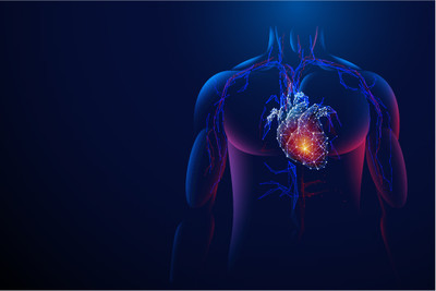
Case study 2: Application of CUT&RUN Technology in Unraveling Epigenetic Mechanisms of Cancer Progression
The CUT&RUN technique operates by employing an antibody to direct micrococcal nuclease (MNase) for the in situ cleavage of chromatin. For professionals in the field, including cancer researchers, oncologists, and pharmaceutical scientists, staying abreast of cutting-edge epigenetic research methodologies is of paramount importance. This technique provides a fresh and invaluable perspective on chromatin analysis in the context of cancer.
- Study Subjects and Enhanced Process
Li and their research team concentrated on cancer cell lines, specifically head and neck cancer cell lines such as HN5, FaDu, and Cal-27. They engineered an enhanced CUT&RUN process that demands low salt concentrations and can function effectively with extremely low cell quantities, as few as 5-20 cells. Additionally, they developed a specialized bioinformatics pipeline named CUT&RUN-Epigenetic-Analyzer (CREA). This process was meticulously crafted to precisely capture the binding sites of key transcription factors implicated in cancer progression, such as p53, NF-κB, and STAT3.
- Superior Performance Compared to Traditional Methods
The outcomes of this study were truly outstanding. The CUT&RUN technique generated high-resolution peaks with a width of less than 100 bp, demonstrating a substantially higher sensitivity compared to the conventional ChIP-seq method. Using just 20 cells, the technique was able to cover 65-95% of the known binding sites of these transcription factors and identify over 800 new co-binding regions involving p53, NF-κB, and STAT3. Moreover, it marked the first instance of accurately quantifying the epigenetic affinity of these cancer-related transcription factors within the native chromatin environment of cancer cells.
- Significance for Cancer Research and Treatment
This optimized CUT&RUN protocol offers a highly efficient and cost-effective tool at the single-nucleotide level for investigating the dynamic regulatory networks of transcription factors in cancer development, progression, and response to therapies. It holds immense potential to propel forward precise epigenetic diagnosis in cancer, enabling the identification of specific epigenetic markers for early detection. Furthermore, it can facilitate the development of targeted epigenetic therapies, which have the potential to revolutionize cancer treatment by specifically modulating the epigenetic alterations that drive cancer progression.
 Application of CUT&RUN in Tumor Research (Womersley et al., 2025)
Frequently Asked Questions Guide
When applying the CUT&RUN technique in practical research, scientists may encounter some common issues. For instance, if there are too few cells and the cell nuclei are not visible, it could be due to excessive cell fixation or incomplete permeabilization, which damages the nuclear structure. In this case, you can appropriately reduce the amount of fixative used or shorten the fixation time. Meanwhile, optimize the permeabilization conditions, such as adjusting the composition of the permeabilization buffer and the permeabilization time.
Industry professionals, including researchers, drug developers, and CROs, need to be well-informed about these practical challenges. Addressing them effectively can ensure the success of CUT&RUN experiments.
1. Recommended Cell Number and Sequencing Depth
Generally, for common transcription factor research, using 50,000-100,000 cells usually yields good experimental results. The sequencing depth depends on the research purpose and genome size. For the human genome, it is recommended to achieve a sequencing depth of 10-20 million reads per sample to accurately detect the binding sites of target proteins.
2. High Background and Solutions
If the analyzed background is too high, in addition to optimizing antibody binding and MNase cleavage conditions as mentioned earlier, you can also consider using negative control samples for the experiment to eliminate non-specific signals in the experimental system. During data analysis, employ appropriate bioinformatics methods to correct and filter out background signals.
3. Cell Samples vs. Nuclear Samples
Whether to use cell samples or nuclear samples for CUT&RUN experiments depends on specific research needs. Cell samples are relatively easy to handle and are suitable for most situations. Nuclear samples, on the other hand, can reduce the interference of cytoplasmic components in the experiment. They may be more advantageous for studying the interactions between nuclear proteins and DNA. For example, when studying the binding sites of certain nuclear transcription factors, using nuclear samples may yield more accurate results.
Conclusion
The CUT&RUN technique, as an emerging epigenetic research method, shows great potential in the field of biological research due to its unique principles and significant advantages. Its characteristics, such as the absence of formaldehyde cross-linking, low background noise, high resolution, and low cell consumption, make it widely applicable in areas like transcription factor binding site analysis and epigenetic modification research.
Through a detailed introduction to the experimental procedure and solutions to common problems, researchers can better master and apply this technique. As research progresses, the CUT&RUN technique is expected to provide more valuable information for revealing gene regulatory mechanisms and the mechanisms of disease occurrence and development, driving the continuous advancement of biological research. In the future, with continuous optimization and innovation, this technique will play an important role in more fields, offering new ideas and methods for solving complex biological problems.
References
- Meers MP, Bryson TD, et al. "Improved CUT&RUN chromatin profiling tools." eLife (2019) 8:e46314
- Miura M, Chen H. "CUT&RUN detects distinct DNA footprints of RNA polymerase II near the transcription start sites." Chromosome Res. 2020;28(3-4):381-393.
- Womersley HJ, Muliaditan D, et al. "Single-nucleus CUT&RUN elucidates the function of intrinsic and genomics-driven epigenetic heterogeneity in head and neck cancer progression." Genome Research. 2025; 35:162-177 .
- Emerson FJ, Lee SS. "CUT&RUN for Chromatin Profiling in Caenorhabditis elegans." Curr. Protoc. 2022;2(6):e445.
- Aflaki S, Margueron R, Holoch D, et al. "Automated CUT & RUN Using the KingFisher Duo Prime." Methods Mol. Biol.2022;2529:253–265.