
 Sample Submission Guidelines
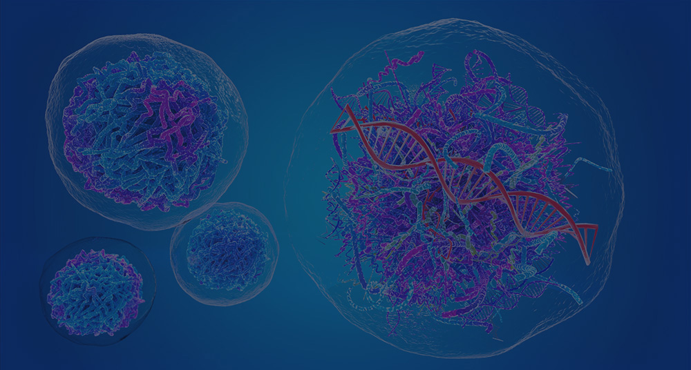
Introduction to SARS-COV-2
In December 2019, the new severe acute respiratory syndrome coronavirus 2 (SARS-CoV-2) started and has enveloped the world in an unparalleled global pandemic. The clinical features of COVID-19 patients are comparable during the 2002-2003 outbreak of SARS-CoV-1, which caused more than 8,000 cases reported and about 800 deaths. As such, human SARS-CoV-2 infection primarily impacts the lower respiratory tract, inducing mild to moderate respiratory symptoms, such as fever, headache, fatigue, myalgia, dry cough, and diarrhea, in about 85% of COVID-19 patients.
SARS-CoV-2 entrance into cells is mediated by a virus surface spike protein. The SARS-CoV-2 spike connects to its human receptor ACE2 (hACE2) through its receptor-binding domain (RBD) to accomplish its purpose and is proteolytically initiated by human proteases. Using biochemical and pseudovirus entry assays, receptor binding was evaluated and protease activation of the SARS-CoV-2 spike. Key cell entrance processes of SARS-CoV-2 were defined by the findings. First, SARS-CoV-2 RBD has a greater affinity for hACE2 binding than SARS-CoV RBD, facilitating the effective entrance of cells. Second, the hACE2 binding affinity of the whole SARS-CoV-2 spike is, paradoxically, similar to or lower than that of the SARS-CoV spike, indicating that SARS-CoV-2 RBD is less vulnerable than SARS-CoV RBD, although more potent. Third, unlike SARS-CoV, proprotein convertase furin preactivated SARS-CoV-2 cell entry, decreasing its reliance on target cell proteases for entrance. SARS-CoV-2 may sustain effective cell entry while avoiding immune surveillance due to the high hACE2 binding affinity of the RBD, furin preactivation of the spike, and hidden RBD in the spike. The widespread of the virus may play a part in these characteristics. Effective intervention techniques must aim at both the potency and the evasiveness of SARS-CoV-2.
Reference Sequence
Causal agent isolation and perseverance of its partial genome sequence given the basis for the SARS-CoV-2 methodologies for viral genome sequencing or real-time reverse transcriptase-polymerase chain reaction (RT-PCR). A diagnostic RT-PCR framework was designed after SARS-CoV-2 was separated from a lower respiratory tract specimen. The RTPCR tests were centered on the ORF1ab sequence RNA-dependent polymerase (RdRp) gene, the SARS-CoV-2 genome’s E gene, N gene, and S gene. RT-PCR assays targeting the RdRp assay had the greatest analytical sensitivity among these assays. In nasal and pharyngeal swab samples, bronchoalveolar lavage fluid, sputum, bronchial aspirates, blood, an anal swab, and other samples, RTPCR may be identified with SARS-CoV-2 nucleic acid. SARS-CoV-2 was instantly identified at the esophageal erosion and at the bleeding site in cases of severe peptic ulcers after the appearance of illness. Gastrointestinal side effects such as diarrhea have also been shown in some patients diagnosed with SARS-CoV-2, as some viruses may access the digestive tract through the throat, contaminate the intestinal epithelial cells and activate the intestinal immune reaction. The nucleic acid SARS-CoV-2 can thus also be identified in some patients’ fecal samples.
For the diagnosis of COVID-19, high-throughput sequencing or an RT-PCR assay has become a normal and formative evaluation. Nucleic acid amplification kits, even so, occasionally yielded false-negative findings among patients whose clinical characteristics, chest imaging, and COVID-19 laboratory identification were consistent. There are many potential explanations for the nuclei acid kit’s false-negative findings. Firstly, even though older age was associated with higher viral load, it is not evident whether there is a positive linear connection between the viral load in body fluids and the intensity of symptoms after infection. If the virus stays quickly replicated and released into the lungs in suspected patients, the sampling of nasal and pharyngeal smears may not obtain sufficient virus for diagnosis. Second, collecting nasal and pharyngeal swabs, sputum, or alveolar lavage fluid is the existing popular sampling technique.
Variant Analysis of SARS-COV-2
The sequence of the established SARS-CoV-2 reference genome, NC 045512, was used in a study conducted last July 2020. In December of 2019, this genome was sequenced. Utilizing the EMBOSS needle (Hinxton, Cambridge, England), each specimen was first positioned pairwise with the reference genome, with a default gap penalty of 10 and an extension penalty of 0.5. In Python (Python Software Foundation, Wilmington, United States of America), this was accompanied by the development of a custom script to obtain the distinctions between the variations of the genome and the reference genome. Nucleotide variants were modified to respective encoded residues of amino acids in the coding areas. The protein coordinates of YP-009724389.1 were utilized for the open reading frame 1 (ORF1) for translation. Finally, stop-gained and frameshift variations have been evaluated that cause deletions and insertions to identify possible artifacts induced by undetermined or ambiguous bases.
References:
- Koyama T, Platt D, Parida L. Variant analysis of SARS-CoV-2 genomes. Bulletin of the World Health Organization. 2020, 98(7).
- Wang C, Liu Z, Chen Z, et al. The establishment of reference sequence for SARS‐CoV‐2 and variation analysis. Journal of medical virology. 2020, 92(6).
- Wang H, Li X, Li T, et al. The genetic sequence, origin, and diagnosis of SARS-CoV-2. European Journal of Clinical Microbiology & Infectious Diseases. 2020.