
Human health and fitness are influenced by the microbial communities that colonize the human gastrointestinal tract. Gut metaproteomic research of whole bacterial diversity give ideas into microorganism phenotypes at the protein rate, and we thank many technological breakthroughs in chromatography and mass spectrometry for our quick advancement.
About Our Structural Proteomics Service
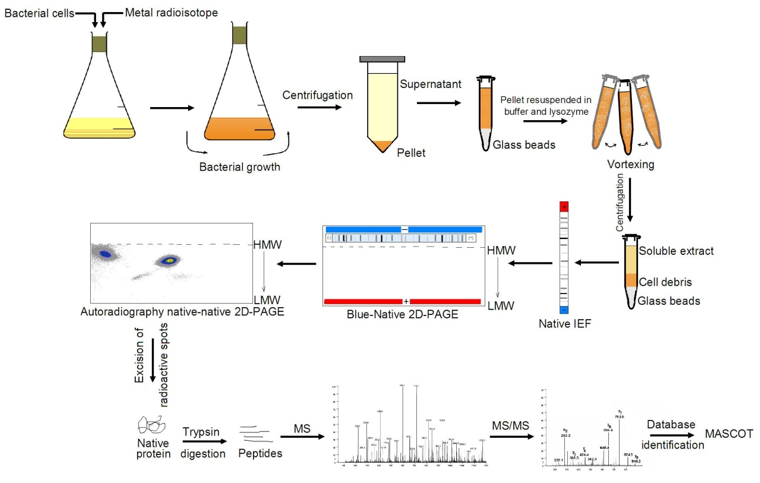
In biological membranes, in complexes, many proteins are coordinated. Through 2D Blue Native/SDS-PAGE analysis, CD Genomics provides a technique for the global assessment of the subunits of these protein complexes. The specimens were evaluated by blue native polyacrylamide gel electrophoresis (BN-PAGE) in the 1st dimension of the 2D BN/SDS-PAGE ass6essment and then divided by SDS-PAGE in the 2nd dimension, which is 90 degrees from the first.
Complex Analysis by 2D Blue Native/SDS-PAGE
The innovation for high-resolution segregation of protein complexes is provided by BN-PAGE. The size, relative abundance, and subunit structure of protein complexes are determined using the blue native electrophoresis procedure. Protein complexes develop and manage the processes of cells and organelles at all rates of time and space complexity, such as development and division of cells, transcription, and translation, breathing and photosynthesis, transport, and metabolism. Protein complexes can show partial and temporal modifications, as well as different reliabilities, making classification and assessment extremely challenging.
Monomeric proteins will relocate in a hyperbolic diagonal due to the gradient gel in the first dimension and a linear gel in the second dimension, while elements of protein complexes will move below this diagonal using a set of first dimension BN-PAGE and second dimension SDS-PAGE. In the second dimension, proteins representing subunits of the same protein complex can be found in a single vertical line, whereas numerous spots of the same protein in the horizontal line imply that the protein is evident in many separate protein complexes.
Our Advantages
- A variety of methodologies for multiple purposes, such as quantification of molecular weight, classification of impurities, research into protein expression, and so on.
- Various specimens, such as fluids from the body, tissues, blood cells, cell cultures, plants, microorganisms, and so on.
- Various sizes of gels to meet different needs.
Workflow and Bioinformatics Analysis

| Bioinformatics Analysis | Details |
| Functional Annotation | Adding sequences of genes or proteins to biological knowledge. To locate similarities the fundamental rate of annotation is to use the sequence alignment device BLAST, and then to annotate genes or proteins depending on that. |
| Functional Enrichment Analysis | Assess gene or protein classes that are over-represented in a huge group of genes or proteins and may have phenotypic relationships with illness. |
| Clustering Analysis | Explore a structure for categorizing observations, such as genes and proteins, into communities with members who share common properties. |
| Network Analysis | Defining the trends of correlation between genes across various specimens |
References
1. Heinemeyer J, Scheibe B, Schmitz UK, Braun HP. Blue native DIGE as a tool for comparative analyses of protein complexes. Journal of proteomics. 2009, 72(3).
2. Klepsch M, Schlegel S, Wickström D, et al. Immobilization of the first dimension in 2D blue native/SDS–PAGE allows the relative quantification of membrane proteomes. Methods. 2008, 46(2).
*For Research Use Only. Not for use in diagnostic procedures.