
Key Aspects of AAV Capsid Protein: Structure, Function, Engineering and Applications
With the rapid development of gene therapy, Adeno-Associated Virus (AAV) has become one of the first choice carriers to deliver therapeutic genes because of its many advantages. In the research of AAV vector development and gene therapy, AAV sequencing has become an indispensable technical support. For example, AAV whole genome sequencing can confirm the integrity and correctness of transgenic sequences. AAV ITR sequence analysis can verify the structural integrity of inverted terminal repeats, and targeted enrichment sequencing (TES) can specifically detect the distribution of AAV vectors in target tissues.
In the whole life cycle of AAV, AAV capsid protein plays a core role. It not only wraps and protects the genome of the virus, but also determines the host range, cytotropism and immunogenicity of the virus. In-depth and comprehensive research on AAV capsid protein is of inestimable significance for optimizing gene therapy strategy, improving therapeutic effect and expanding the application scope of gene therapy.
This paper focuses on the capsid protein of AAV, and expounds its structural characteristics, functions, engineering techniques, analytical methods and applications.
AAV Capsid Protein Characteristics
AAV, as a non-pathogenic single-stranded DNA virus, shows great potential in gene therapy and vaccine development, in which AAV capsid protein plays a key role.
Structure of Capsid Protein Subunit
AAV capsid proteins are mainly composed of three different subunits, namely VP1, VP2 and VP3, and their ratio in virus particles is about 1:1:10. These three subunits have similar core domains, but there are differences in N-terminal and C-terminal.
Core domain: The core domains of VP1, VP2 and VP3 all contain multiple β-sheet and α-helix structures. These secondary structural elements interact with each other through a specific connection mode, forming a tightly stacked three-dimensional structure. Core domain is very important to maintain the overall stability of capsid, and also participates in the initial recognition and binding process between virus and host cell receptor.
N-terminal domain: The N-terminal domain of VP1 is relatively long and contains a unique phospholipase A2 (PLA2) domain. This domain plays an important role in the process of virus infection. It can hydrolyze phospholipids on the host cell membrane, promote the fusion of virus and cell membrane, and thus assist the virus genome to enter the host cell. The N-terminal domain of VP2 is short, which is similar to the N-terminal sequence of VP1, but lacks the complete PLA2 active domain. VP3 has almost no obvious N-terminal extension domain.
C-terminal domain: The C-terminal domains of the three subunits are also different in length and amino acid sequence. C-terminal domain is involved in the interaction between capsid protein subunits, which has an important influence on the assembly and stability of capsid. In addition, there are some key amino acid residues in the C-terminal domain, which may be involved in the interaction between the virus and host intracellular factors, affecting the infection efficiency and tissue tropism of the virus.
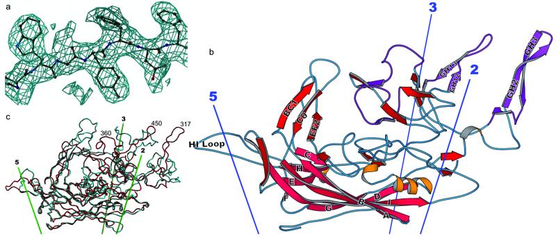 Structure of the AAV-2 subunit and comparison with related structures (Xie et al., 2002)
Surface Structure of Capsid Protein
The surface of AAV capsid is not smooth, but has a series of unique structural features, which are closely related to the infection characteristics of virus.
Spine: On the surface of AAV capsid, there are some protruding spines. These spikes are composed of specific regions of capsid protein subunits, which are usually located at the apex or edge of icosahedron. Spine structure plays a key role in the binding process between virus and host cell receptor. The structure and amino acid composition of spines of different serotypes of AAV are different, which also leads to their different recognition specificity for different host cell receptors, and then determines the tissue tropism of AAV.
Depressions: In addition to spikes, there are some depressed areas on the shell surface. These depressions may be the sites where viruses interact with helper receptors or other cytokines on the surface of host cells. Studies have shown that some small molecules or antibodies can bind to the concave areas on the capsid surface, thus affecting the virus infection process, which provides a potential target for developing antiviral strategies against AAV.
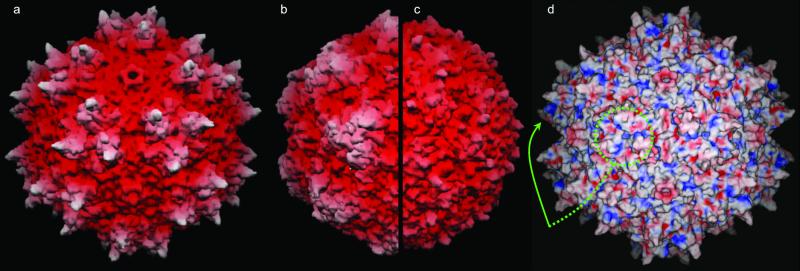 Surface topology and electrostatics (Xie et al., 2002)
How Does AAV Capsid Protein Function
The primary and basic function of capsid protein is to protect the genetic material of virus. The genome of AAV is single-stranded DNA, which is easily damaged by nucleases and other factors in the extracellular environment. By tightly wrapping DNA, capsid protein provides a physical barrier to prevent the degradation of nuclease, and ensures the integrity and stability of virus genome in the process of transmission.
Capsid protein plays a key role in mediating the recognition and combination of virus and host cells. The capsid surface has specific domains and amino acid sequences, which determine the tendency of AAV to different host cell types.
After the virus enters the host cell, the capsid protein also participates in the release process of the virus genome. When AAV is endocytosed, it will undergo a series of intracellular transport processes and reach the vicinity of the nucleus. In this process, the conformation of capsid protein will change. The conformation-changed capsid protein can help the virus genome to be released from the capsid, so that it can enter the nucleus and start the transcription and replication of virus genes, thus completing the infection cycle of the virus.
In addition, AAV capsid protein also has an important influence on the immunogenicity of the virus. The recognition and response of the immune system to the virus largely depends on the structure and composition of capsid protein. Although AAV is generally regarded as a virus vector with relatively low immunogenicity, capsid protein may still trigger the host's immune response.
On the one hand, the immune system can recognize some epitopes of capsid protein and trigger immune response, which may affect the distribution and persistence of virus vectors in the body. By modifying or transforming capsid protein, the immunogenicity of the virus can be adjusted, and its safety and effectiveness as a gene therapy vector can be improved.
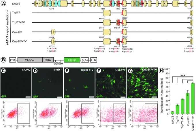 Scale representation of the AAV2 capsid (Cap) protein shared VP1/VP2/VP3 C terminus highlighting the position of each capsid mutation (Lipinski et al., 2015)
Take the Next Step: Explore Related Services
Learn More
- AAV in Gene Therapy: Rationale, Mechanisms, Applications, Safety, and Efficiency Enhancement
- In-Depth Exploration of AAV: From Biological Traits to Gene Therapy Horizons
- AAV ITR Sequencing: Unraveling the Workflow, Result Analysis, Technical Hurdles, and Future Trends
- Viral Therapy Explained: Concepts, Classifications, and Clinical Applications
AAV Capsid Protein Engineering
Accurate engineering transformation and modification of capsid protein can not only deeply understand the biological characteristics of virus, but also provide broad application prospects for developing new antiviral strategies, gene therapy vectors and vaccines.
Site-directed Mutation Technique
Site-directed mutation is a technique to precisely change the specific amino acid residues of capsid protein. By designing primers with specific mutation sites, the gene fragment encoding capsid protein in virus genome was amplified by PCR technology, and the mutation was introduced into the target gene. Then, the mutated gene was cloned into an appropriate expression vector, expressed in the host cell and assembled into virus particles with mutated capsid protein.
Site-directed mutation can be used to study the role of specific amino acid residues in the structure and function of capsid protein. In addition, site-directed mutation can also be used to improve the performance of viral vectors, such as enhancing the targeting of vectors or reducing their immunogenicity.
Gene Fusion Technology
Gene fusion technology is to fuse the gene encoding capsid protein with other functional genes, so that capsid protein can carry additional functional domains when expressed. This fusion protein can endow virus particles with new characteristics. The green fluorescent protein (GFP) gene was fused with the virus capsid protein gene, and the expressed capsid protein was labeled with fluorescence, which was convenient for observing the infection process and distribution of the virus in the host cell under the microscope. In addition, the fusion of targeted polypeptide gene and capsid protein gene can make virus particles specifically target specific tissues or cell types, and improve the targeting of virus vectors in gene therapy.
Chemical Modification Method
Chemical modification is the chemical treatment of purified capsid protein or virus particles in vitro to change their chemical structure and properties. Common chemical modification methods include glycosylation modification, PEGylation modification and lipidation modification. Glycosylation modification can change the surface charge and antigenicity of virus particles by adding sugar chains on the surface of capsid protein, and affect the interaction between virus and host cells and the recognition of virus by host immune system.
Pegylation modification is to covalently attach polyethylene glycol (PEG) to capsid protein, which increases the water solubility and stability of virus particles, reduces their immunogenicity and prolongs the circulation time of virus vectors in vivo. Lipid modification is to connect lipid molecules to capsid protein, which makes it easier for virus particles to interact with cell membrane and improve the infection efficiency of virus.
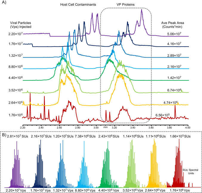 ZipChip CE-MS Limit of Detection testing for AAV capsid analysis (Smith et al., 2024)
AAV Capsid Protein Characterization Analysis
Capsid protein plays a key role in the structural integrity, infectivity and host immune response induction of virus. Understanding the characteristics of capsid protein is very important for explaining the life cycle of virus, developing antiviral strategies and designing biological products based on virus.
Post-translation Modification Analysis
Capsid proteins often undergo various post-translational modifications, such as phosphorylation and glycosylation, which affect the function of proteins. Mass spectrometry, such as liquid chromatography-mass spectrometry (LC-MS/MS), can identify the modification sites and types by detecting the mass changes caused by modification. In the study of herpes virus, it is found that the phosphorylation of capsid protein regulates its interaction with host cell proteins and affects the replication and spread of the virus.
Ligand Binding Assay
Studying the binding characteristics of capsid protein with other molecules (such as receptors and nucleic acids) is helpful to understand the mechanism of virus infection. Surface plasmon resonance (SPR) technology can monitor the interaction between molecules in real time, and accurately measure the binding affinity and dissociation constant by detecting the change of resonance signal when proteins bind to ligands. In the study of influenza virus, the binding kinetics of capsid protein with sialic acid receptor on the surface of host cells was analyzed by SPR, which provided the target basis for the design of anti-influenza drugs.
Viral Infectivity Assay
It is the key of functional analysis to directly evaluate the effect of capsid protein on virus infectivity. By constructing capsid protein mutant, the infection titer and host range of recombinant virus were determined. In HIV research, the specific domain of capsid protein was mutated to observe its influence on the ability of HIV to infect host T cells, revealing the key function of capsid protein in the process of virus entry, shelling and reverse transcription.
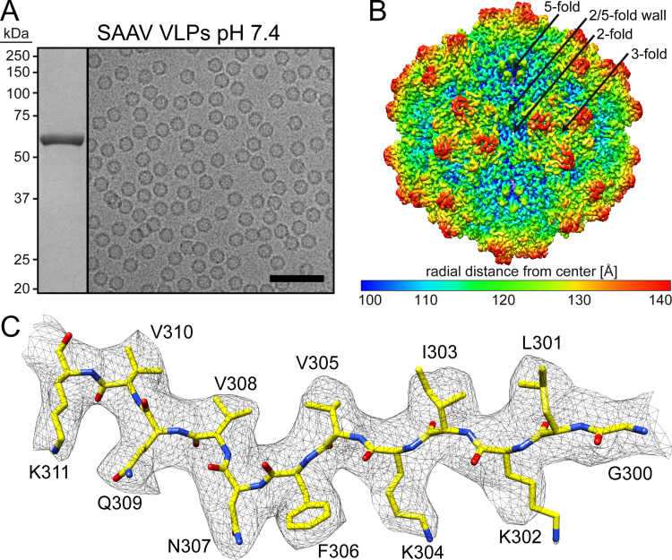 SAAV VLPs cryo-EM structure (Mietzsch et al., 2022)
AAV Capsid Protein Application
The unique structure and properties of AAV capsid protein endow AAV carriers with diverse application potential. The in-depth study on the application of AAV capsid protein not only promotes the development of cutting-edge medical technologies such as gene therapy, but also brings innovative solutions to the field of bioengineering.
Application of Gene Therapy Vector
AAV capsid protein can efficiently wrap foreign genes and deliver them to target cells accurately. AAV vector has shown great advantages in the treatment and research of many monogenic diseases.
By modifying AAV capsid protein, targeted gene delivery to specific tissues or cell types can be realized. Using phage display technology, the researchers fused specific peptide fragments to the surface of AAV capsid protein, and constructed AAV vectors with liver targeting.
Development and Application of Vaccine
AAV capsid proteins can self-assemble to form virus-like particles (VLPs). These VLPs do not contain the genome of the virus, so they are not infectious, but they retain the natural antigenic epitopes of the virus. In the development of vaccine, VLP based on AAV capsid protein can effectively activate the immune system and produce specific immune response. Compared with traditional vaccine, VLP vaccine based on AAV capsid protein has better safety and immunogenicity, and is easy to produce on a large scale. AAV capsid protein can also be used as a carrier to deliver immunomodulatory molecules, which can be used to treat immune-related diseases or enhance the immune effect of vaccines.
Construction of Biological Nano-materials
AAV capsid protein has a highly ordered structure and good biocompatibility, and can be used as an ideal template for constructing biological nanomaterials. Researchers have successfully prepared biological nanocomposites with specific functions by modifying functional groups, such as metal ion binding sites, on the surface of AAV capsid protein. These nano-materials have potential application value in the fields of biological imaging, drug delivery and catalysis.
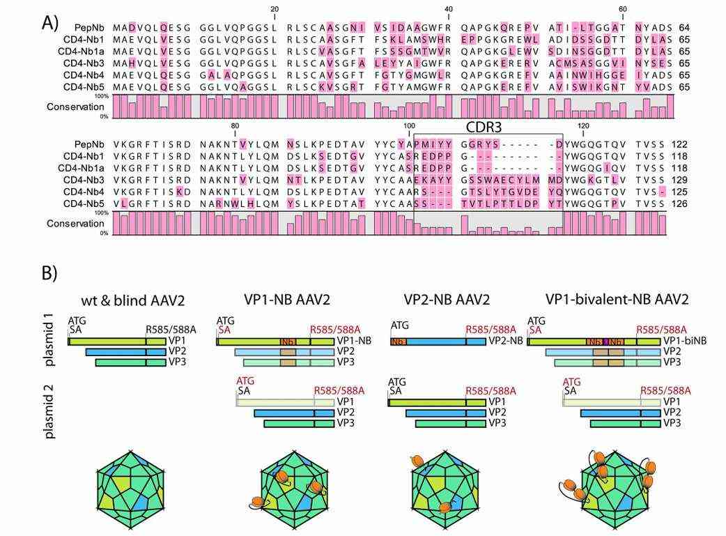 (Nb) sequences and AAV2-Nb design (Hamann et al., 2021)
Conclusion
AAV capsid protein shows unparalleled advantages in the field of gene therapy, and its unique structure endows the virus with high stability and tissue targeting specificity. However, the research of AAV capsid protein still faces many challenges, such as the regulation of immunogenicity and the construction of efficient targeted delivery system. However, with the rapid development of science and technology, emerging technologies are constantly emerging, and the research on AAV capsid protein is expected to make a major breakthrough in the future.
References
- Xie Q, Bu W., et al. "The atomic structure of adeno-associated virus (AAV-2), a vector for human gene therapy." Proc Natl Acad Sci U S A. 2002 99(16):10405-10 https://doi.org/10.1073/pnas.162250899
- Mietzsch M, Hull JA., et al. "Characterization of the Serpentine Adeno-Associated Virus (SAAV) Capsid Structure: Receptor Interactions and Antigenicity." J Virol. 2022 96(11):e0033522 https://doi.org/10.1128/jvi.00335-22
- Lipinski DM, Reid CA., et al. "Systemic Vascular Transduction by Capsid Mutant Adeno-Associated Virus After Intravenous Injection." Hum Gene Ther. 2015 26(11):767-76 https://doi.org/10.1089/hum.2015.097
- Martin V. Hamann , Niklas Beschorner., et al. "Improved targeting of human CD4 by nanobody-modified AAV2gene therapy vectors." PLoS ONE. 2021 16(12): e0261269 https://doi.org/10.1371/journal.pone.0261269
- Smith J, Carillo S., et al. "Rapid characterization of adeno-associated virus (AAV) capsid proteins using microchip ZipChip CE-MS." Anal Bioanal Chem. 2024 416(4):1069-1084 https://doi.org/10.1007/s00216-023-05097-5
- Zhang Z, Park J., et al. "Capillary Electrophoresis-Sodium Dodecyl Sulfate with Laser-Induced Fluorescence Detection as a Highly Sensitive and Quality Control-Friendly Method for Monitoring Adeno-Associated Virus Capsid Protein Purity." Hum Gene Ther. 2021 32(11-12):628-637 https://doi.org/10.1089/hum.2020.233


