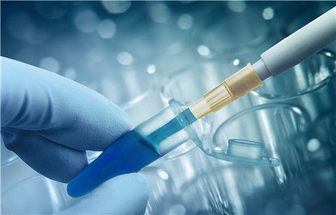

Tumor Organoid–Immune Co-Culture: Study Design and Validation

Designing a tumor organoid–immune co-culture experiment requires balancing biological fidelity, cellular viability, and technical reproducibility. Standard two-dimensional cell lines fail to replicate the structural heterogeneity and spatial architecture of solid tumors, while in vivo animal models often present cross-species immunological discrepancies and long experimental turnaround times. Three-dimensional patient-derived organoids (PDOs) combined with functional immune compartments bridge this gap, enabling ex vivo investigation of antigen presentation, immune cell infiltration, tumor killing kinetics, and immunotherapy response mechanisms.
This guide provides a practical framework for planning tumor organoid–immune co-culture studies, from model and immune cell selection to experimental controls, functional readouts, and downstream molecular validation.
Disclosure: CD Genomics provides specialized research-use-only (RUO) organoid multi-omics solutions, organoid single-cell RNA sequencing, and TCR sequencing to support preclinical discovery and translational research; our services are strictly intended for scientific research purposes and do not provide clinical diagnostic or therapeutic guidance.
TL;DR
- Model selection dictates analytical depth: Choose holistic native platforms (such as air-liquid interface cultures or precision-cut tumor slices) when preserving endogenous tumor-infiltrating lymphocytes (TILs) and native stroma for short-term assays (≤8–14 days) is critical. Choose reconstituted reductionist platforms (passaged epithelial PDOs re-seeded with exogenous autologous PBMCs or engineered T/NK cells) when modularity, scalability, and long-term reproducibility are needed.
- Autologous sourcing eliminates alloreactivity: Testing physiological tumor antigen recognition requires autologous immune effectors (PBMCs or TILs) matched to patient organoids. Allogeneic effectors introduce major histocompatibility complex (MHC) mismatch artifacts, requiring unmanipulated baseline controls to distinguish alloreactivity from tumor-antigen-specific cytotoxicity.
- Multi-modal endpoints provide complete answers: Pairing live-cell cytotoxicity imaging with multiplex cytokine profiling, flow cytometry, single-cell RNA sequencing (scRNA-seq), and T-cell receptor (TCR) sequencing enables researchers to correlate physical tumor killing with cell-state transitions and antigen-specific clonotype expansion.
If your team is currently establishing broader organoid sequencing protocols, consult our foundational organoid sequencing study design guide.
Model Architecture: Native vs Reconstituted Co-Culture Systems
Selecting an appropriate culture architecture is the primary decision in study design. The chosen system determines which cellular compartments are preserved, how long cells remain viable, and which perturbation assays can be executed reliably.
Holistic Native Models (Air-Liquid Interface and Tumor Slices)
Holistic co-culture platforms preserve the native tumor microenvironment (TME) directly from fresh surgical resections or core biopsies without enzymatic dissociation into single cells. In the Air-Liquid Interface (ALI) method, mechanically diced primary tumor fragments embedded in a type I collagen matrix are cultured on a semi-permeable transwell insert, exposing the top of the collagen gel to air while nutrients diffuse from the culture medium below (Neal et al., 2018, Cell: Organoid Modeling of the Tumor Immune Microenvironment).
ALI cultures retain endogenous tumor parenchymal cells alongside tumor-infiltrating lymphocytes (CD4+ and CD8+ T cells, B cells), myeloid-derived suppressor cells (MDSCs), tumor-associated macrophages (TAMs), and cancer-associated fibroblasts (CAFs). This endogenous preservation allows immediate evaluation of immune checkpoint inhibitors without requiring in vitro immune cell expansion. However, endogenous immune cell populations decline steadily ex vivo, typically limiting experimental viability windows to 7–14 days (Magré et al., 2023; Voabil et al., 2021, Nature Medicine: An ex vivo tumor fragment platform). Furthermore, non-dissociated fragments display considerable inter-well variability in size and cellular composition, preventing high-throughput miniaturization.
Reconstituted Reductionist Models (Extracellular Matrix Hydrogels)
The reconstituted approach decouples tumor organoid propagation from immune cell isolation. Primary tumor tissues are enzymatically dissociated to generate pure epithelial PDO lines embedded in basement membrane extract (BME or Matrigel). Once established, mature PDOs are harvested, fragmented or dissociated, and co-cultured with separately isolated and characterized immune effector populations (Cattaneo et al., 2020, Nature Protocols: Tumor organoid–T-cell coculture systems; Dijkstra et al., 2018, Cell: Generation of Tumor-Reactive T Cells).
Reconstituted models provide defined, modular control over experimental variables. Researchers can titrate specific effector-to-target (E:T) ratios, compare distinct immune subsets (e.g., purified CD8+ T cells vs. total PBMCs vs. NK cells), and perform genetic perturbations in the organoid or immune compartment independently. The trade-off is that reconstituted systems lack native stromal architecture unless CAFs or endothelial cells are explicitly co-seeded. Furthermore, standard organoid expansion conditions may contain components that alter immune cell phenotype or suppress immune activation, so culture compatibility should be evaluated before immune cell addition (Magré et al., 2023).
Microfluidic and Organoid-on-a-Chip Systems
Organoid-on-a-chip platforms integrate 3D microfluidic channels with hydrogel-embedded organoids and endothelialized barriers. These microphysiological systems model continuous interstitial fluid flow, nutrient and oxygen gradients, and dynamic immune cell extravasation from vascular channels into the tumor compartment (Wang et al., 2025, Cell Death Discovery: Tumor organoid-immune co-culture models). While they provide high spatiotemporal resolution for tracking immune cell migration and infiltration kinetics, they require specialized microfabrication equipment and offer lower throughput than standard microtiter plate formats.
Structural Comparison of Co-Culture Architectures
| Parameter | Holistic Native (ALI / Tumor Slices) | Reconstituted Reductionist (PDO + Immune Cells) | Microfluidic Organoid-on-a-Chip |
|---|---|---|---|
| Cellular Composition | Native tumor, endogenous TILs, myeloid cells, CAFs | Epithelial PDOs + defined exogenous immune subsets | Epithelial PDOs + immune cells ± endothelial cells |
| TME Fidelity | High (native spatial architecture preserved) | Moderate (reconstructed modular architecture) | High (dynamic fluidic shear stress & vascular interface) |
| Culture Longevity | Short-term (typically 7–14 days) | Medium-to-long (weeks; PDOs passaged indefinitely) | Short-to-medium (typically 3–10 days) |
| Throughput Potential | Low to moderate (6- to 24-well transwells) | High (96- to 384-well plate formats) | Low (specialized microfluidic chips) |
| Primary Use Case | Rapid ex vivo checkpoint blockade screening | Scalable T/NK cytotoxicity, CAR testing, MoA studies | Infiltration kinetics, extravasation, vascular biology |
 Figure 2. Structural and biological comparison of holistic native, reconstituted reductionist, and organoid-on-a-chip co-culture platforms.
Immune Cell Sourcing: Autologous vs Non-Autologous Strategies
The choice of immune effector source directly influences the immunological questions a study can answer. Distinguishing tumor-antigen-restricted cytotoxicity from allogeneic graft-versus-tumor effects is critical when selecting cell sources.
Autologous Peripheral Blood Mononuclear Cells (PBMCs)
Autologous PBMCs isolated from patient blood drawn at the time of tumor biopsy or surgery represent a readily accessible, non-destructive immune cell source. Because they share the patient's HLA haplotype, autologous PBMCs eliminate allogeneic background responses (Dijkstra et al., 2018; Magré et al., 2023, JITC: Emerging organoid-immune co-culture models).
Circulating tumor-reactive T cells are typically present at low frequencies in peripheral blood. Successful co-culture protocols utilize repeated cycles of co-culture stimulation (typically 1–3 rounds over 10–21 days in the presence of low-dose IL-2 and IL-15) with matched tumor organoids to selectively expand tumor-reactive CD8+ T cells (Cattaneo et al., 2020; Dijkstra et al., 2018). This setup is best suited for evaluating neoantigen-specific T-cell priming, memory recall, and peripheral immune repertoire mobilization.
Autologous Tumor-Infiltrating Lymphocytes (TILs)
TILs extracted directly from digested primary tumor tissue are pre-enriched for tumor-antigen-experienced T-cell clones. Freshly isolated TILs frequently display an exhausted or dysfunctional phenotype marked by high surface expression of PD-1, TIM-3, LAG-3, and CTLA-4 (Jeong & Kang, 2023, IJMS: Exploring Tumor–Immune Interactions in Co-Culture Models; Zhao et al., 2023, Cells: Organoids as an Enabler of Precision Immuno-Oncology). Co-culture of TILs with matched PDOs serves as an ideal baseline model for evaluating whether immune checkpoint inhibitors or costimulatory agonists (e.g., 4-1BB/CD137 agonists) can reverse exhaustion and restore cytotoxic killing.
Innate Effectors: Allogeneic NK Cells and γδ T Cells
When autologous patient material is limited or when evaluating off-the-shelf cell therapies, innate effectors such as natural killer (NK) cells or gamma-delta (γδ) T cells are commonly employed (Wang et al., 2025; Zhao et al., 2023). NK cells and γδ T cells recognize stress-induced ligands (e.g., MICA/MICB, ULBPs) and metabolic intermediates on transformed epithelial cells independently of classical MHC-I presentation. This allows them to eliminate tumor organoids that have down-regulated HLA machinery to escape CD8+ T cells. When utilizing allogeneic primary NK cells or expanded αβ T cells, researchers must include matched normal epithelial organoids (derived from healthy adjacent tissue) to quantify baseline allogeneic lysis.
Immune Sourcing Decision Table
| Immune Cell Source | HLA Compatibility | Antigen Experience | Expansion Requirement | Primary Application |
|---|---|---|---|---|
| Autologous PBMCs | 100% matched | Low frequency in circulation | Requires 1–3 rounds of organoid stimulation | TCR clone discovery, neoantigen reactivity, T-cell priming |
| Autologous TILs | 100% matched | High (tumor-antigen experienced) | Limited yield; short-term or rapid expansion protocol | Checkpoint blockade rescue, exhaustion reversal, adoptive cell therapy pre-testing |
| Allogeneic NK Cells | Mismatched (MHC-independent) | Innate activation by stress ligands | Feasible off-the-shelf expansion | Non-MHC restricted tumor killing, ADCC assays, CAR-NK validation |
| Engineered CAR-T / TCR-T | Autologous or Allogeneic | Defined synthetic specificity | Pre-engineered viral/non-viral transduction | On-target cytotoxicity, off-tumor safety, target density thresholds |
Experimental Variables, Controls, and Timeline Optimization
Achieving reproducible data in tumor organoid–immune co-cultures requires rigorous calibration of experimental kinetics, medium formulations, effector ratios, and control arms.
Effector-to-Target (E:T) Ratio Optimization
The ratio of immune effector cells to organoid cells must be titrated empirically. In 3D Matrigel dome co-cultures, excessive effector numbers can induce rapid, non-specific physical degradation of the extracellular matrix, whereas insufficient ratios yield undetectable killing signals.
- Purified CD8+ T Cells / Expanded TILs: Effective ratios typically range from 1:1 to 5:1 (effectors:organoid cells).
- Unselected Total PBMCs: Typical starting ranges are 10:1 to 20:1, accounting for the lower percentage of CD3+ T cells and antigen-specific clones within total mononuclear populations.
- Engineered CAR-T / NK Cells: Potent effector constructs typically operate at ratios between 0.5:1 and 2:1.
Medium Harmonization and Compatibility Assessment
One of the most frequent points of failure in reconstituted co-cultures is medium incompatibility. Standard organoid expansion medium and immune cell culture medium contain conflicting components:
- Expansion-Medium Compatibility: Some organoid culture supplements can interfere with immune cell proliferation or cytokine release. Their compatibility should be evaluated and adjusted using a validated co-culture protocol (Magré et al., 2023).
- Culture Additives: Small-molecule supplements used during organoid expansion may influence immune signaling and should be reviewed for compatibility before immune cell addition.
- Cytokine Supplementation: Co-culture media should be supplemented with low-dose IL-2 (20–100 IU/mL) and IL-15 (5–10 ng/mL) to maintain immune viability without inducing non-specific, antigen-independent T-cell activation.
Essential Experimental Groups and Controls
Every robust co-culture design requires structured control arms to isolate specific immune-mediated mechanisms from background matrix loss or non-specific compound toxicity:
- Group 1 (Organoid Alone): Baseline organoid viability and spontaneous death rate over the culture timeframe.
- Group 2 (Organoid + Isotype Ab): Baseline antibody-dependent background and non-specific binding controls.
- Group 3 (Organoid + Immune Effectors + Isotype Ab): Basal antigen-specific killing and baseline alloreactivity.
- Group 4 (Organoid + Immune Effectors + Checkpoint Inhibitor): Treatment-induced immune reactivation and cytotoxicity.
- Group 5 (Matched Normal Organoid + Immune Effectors): Safety benchmark generated from non-cancerous adjacent tissue to confirm that cytotoxicity is tumor-specific and not driven by general epithelial lysis (Cattaneo et al., 2020; Dijkstra et al., 2018).
- Group 6 (Immune Alone + Treatment): Baseline immune activation and background cytokine release in the absence of target cells.
Pre-incubating tumor organoids with anti-HLA-A/B/C blocking antibodies (clone W6/32) prior to T-cell addition is also recommended to verify whether observed killing is strictly canonical MHC class I-dependent (Dijkstra et al., 2018).
Multi-Modal Readouts: From Phenotypic Killing to Single-Cell Omics
To capture the full spectrum of tumor–immune interaction, studies should combine functional killing assays with molecular, phenotypic, and genomic profiling.
Functional Cytotoxicity and Real-Time Imaging
Static end-point assays often fail to capture the dynamic kinetics of immune attack. Integrating live-cell confocal or high-content imaging provides time-resolved spatial data. Cell-permeable fluorogenic caspase substrates (e.g., NucView 488) added to the co-culture generate green fluorescent signals exclusively upon apoptosis induction inside organoids, enabling real-time quantification of apoptotic area over 24–96 hours. Pre-labeling organoids with stable cytoplasmic dyes (e.g., CellTrace Far Red or Calcein-AM) while tracking immune effectors with distinct fluorophores allows automated image segmentation algorithms to differentiate organoid disintegration from immune cell death.
Secretome and Cytokine Profiling
Supernatants collected at defined intervals (e.g., 24, 48, and 72 hours) reflect the activation state and effector mechanism of the immune compartment. Multiplex bead-based immunoassays (Luminex) or ELISAs should quantify IFN-γ, TNF-α, Granzyme B, and Perforin to confirm cytotoxic degranulation. Concurrently, measuring IL-6, IL-10, TGF-β, CXCL9, and CXCL10 helps characterize TME chemokine gradients and feedback inhibition loops.
Flow Cytometry Phenotyping
Harvesting the non-adherent and matrix-dissociated cell fractions allows detailed surface and intracellular marker quantification, including CD3, CD4, CD8, CD25, CD69, CD137 (4-1BB), PD-1, TIM-3, LAG-3, and TIGIT for T cells, CD107a (LAMP-1) for degranulation, and HLA-ABC, HLA-DR, β2-microglobulin (B2M), and PD-L1 expression on epithelial tumor cells (EpCAM+).
High-Resolution Single-Cell RNA Sequencing (scRNA-seq)
While bulk transcriptomics averages signals across all cell types, organoid single-cell RNA sequencing resolves the distinct transcriptional responses of individual tumor and immune sub-populations simultaneously (Jeong & Kang, 2023; Magré et al., 2023):
- Tumor Heterogeneity & Evasion Programs: Identify specific tumor sub-clones that upregulate interferon-stimulated genes (ISGs) versus those that down-regulate antigen-processing machinery (TAP1/2, B2M) under immune pressure.
- Ligand-Receptor Interactome Analysis: Bioinformatic tools (such as CellChat or SingleCellSignalR) applied to co-culture scRNA-seq datasets infer active communication channels between cancer cells and immune subsets (e.g., CD274–PDCD1, LGALS9–HAVCR2, PVR–TIGIT).
T-Cell Receptor (TCR) Repertoire Sequencing
Coupling co-culture experiments with deep TCR sequencing enables tracking of clonal dynamics before and after organoid stimulation (Jeong & Kang, 2023). Researchers can identify specific CDR3 sequences that expand selectively in response to tumor organoids but not normal organoids, calculate Shannon entropy and Simpson clonality, and track oligoclonal convergence to discover candidate tumor-reactive TCRs for downstream transgenic expression. For advanced context on how lymphoid structures drive immune diversity in tissue niches, see our review on tertiary lymphoid structures in immunotherapy research.
 Figure 3. Integrative readout matrix connecting phenotypic functional assays with high-dimensional single-cell transcriptomics and TCR clonotype profiling.
Research Question to Readout Matrix
| Research Objective | Optimal Co-Culture Architecture | Recommended Immune Sourcing | Primary Readout Stack | Key Validation Control |
|---|---|---|---|---|
| Ex Vivo Checkpoint Efficacy (anti-PD-1/PD-L1) | Holistic Native (ALI / Tumor Slices) | Endogenous TILs + TME stroma | Secreted IFN-γ/TNF-α, live/dead imaging, IHC | Matched isotype control antibody |
| Tumor-Reactive T-Cell Discovery & Priming | Reconstituted 3D Matrigel PDOs | Autologous PBMCs (multi-round co-culture) | TCR sequencing, CD137 upregulation, IFN-γ ELISpot | Matched normal organoids (unstimulated) |
| CAR-T / CAR-NK Specificity & Off-Target Lysis | Reconstituted 3D PDOs | Engineered CAR-T or CAR-NK cells | Real-time Caspase-3/7 imaging, LDH release, Granzyme B | Non-transduced T cells & antigen-negative organoids |
| TME Infiltration & Chemotaxis Dynamics | Microfluidic Organoid-on-a-Chip | Autologous or Allogeneic T/NK cells | Time-lapse confocal microscopy, spatial tracking | Non-chemotactic negative control channel |
| Immune Escape & Cell-State Transition Profiling | Reconstituted 3D PDOs | Autologous TILs or primed PBMCs | Organoid single-cell RNA-seq + TME profiling | Untreated organoid and immune baseline cohorts |
Data Interpretation Boundaries and Downstream Molecular Validation
While tumor organoid–immune co-cultures offer biological relevance, investigators must account for specific in vitro boundaries when interpreting findings for translational programs.
Methodological Limitations to Consider
Dense reconstituted basement membrane matrices (e.g., high-concentration Matrigel domes) can physically impede T-cell migration and direct cell-to-cell contact, occasionally underestimating immune potency compared to suspension or low-percentage hydrogel systems (Jeong & Kang, 2023; Magré et al., 2023). Furthermore, standard static 3D organoids lack continuous blood perfusion, endothelial shear stress, and lymphatic drainage; systemic pharmacokinetic behavior and distant homing kinetics cannot be modeled in static well plates. Finally, prolonged culture in high-concentration recombinant cytokines or continuous exposure to high tumor burdens can induce culture-induced exhaustion phenotypes that differ from in vivo exhaustion states (Magré et al., 2023; Wang et al., 2025).
Orthogonal Validation Strategies
To confirm that co-culture discoveries reflect true biological mechanisms rather than in vitro artifacts, project teams should establish an orthogonal validation framework:
- Target Dependency via Functional Genetics: If a co-culture screen identifies a putative immune-sensitizing target or resistance pathway, validate the mechanism by knocking out or overexpressing the candidate gene in the organoid line prior to repeat co-culture.
- Spatial and Orthogonal Tissue Confirmation: Cross-reference organoid multi-omics signatures against spatial transcriptomics or multiplex immunofluorescence (mIF) staining of the original primary patient tumor tissue blocks.
- Preclinical In Vivo Benchmarking: Candidate immunotherapy regimens or expanded tumor-reactive T-cell products identified in organoid co-cultures can be functionally validated in syngeneic murine models or humanized patient-derived xenograft (CDX/PDX) models where systemic immunological organs are intact.
 Figure 4. Decision and validation framework translating ex vivo organoid-immune co-culture findings to preclinical models and spatial validation.
Partnering with CD Genomics for Advanced Preclinical Profiling
Accelerating immuno-oncology discovery requires integrated workflows spanning organoid model characterization, immune profiling, and deep molecular analysis. CD Genomics offers end-to-end preclinical support through dedicated immuno-oncology solutions, comprehensive tumor microenvironment profiling, and high-resolution TCR sequencing in immune research. All our laboratory and bioinformatics services are strictly designed for research use only (RUO) to empower biopharma and academic discovery teams with reliable, publication-ready multi-omics data.
FAQ
- Q1. What is the primary difference between native and reconstituted tumor organoid co-cultures?
- Q2. Why is medium formulation critical in organoid–immune co-culture experiments?
- Q3. How do researchers differentiate tumor-specific killing from allogeneic background lysis?
- Q4. What effector-to-target (E:T) ratio should be used for initial co-culture optimization?
- Q5. What are the main limitations of static 3D organoid co-cultures in drug discovery?
- Q6. How does single-cell RNA sequencing add value to organoid co-culture studies?
More practical guides and application notes are available in the Biomedical NGS Learning Center.
References
- Magré L, Verstegen MMA, Buschow SI, van der Laan LJW, Peppelenbosch MP, Desai J. Emerging organoid-immune co-culture models for cancer research: from oncoimmunology to personalized immunotherapies. Journal for ImmunoTherapy of Cancer. 2023;11(5):e006290. DOI: 10.1136/jitc-2022-006290
- Jeong SR, Kang M. Exploring Tumor–Immune Interactions in Co-Culture Models of T Cells and Tumor Organoids Derived from Patients. International Journal of Molecular Sciences. 2023;24(19):14609. DOI: 10.3390/ijms241914609
- Zhao J, Fong A, Seow SV, Toh HC. Organoids as an Enabler of Precision Immuno-Oncology. Cells. 2023;12(8):1165. DOI: 10.3390/cells12081165
- Wang J, Tao X, Zhu J, Dai Z, Du Y, Xie Y, Chu X, Fu G, Lei Z. Tumor organoid-immune co-culture models: exploring a new perspective of tumor immunity. Cell Death Discovery. 2025;11:195. DOI: 10.1038/s41420-025-02407-x
- [5] Voabil P, de Bruijn M, Roelofsen LM, Hendriks SH, Brokamp S, van den Braber M, et al., Thommen DS. An ex vivo tumor fragment platform to dissect response to PD-1 blockade in cancer. Nature Medicine. 2021;27(7):1250-1261. DOI: 10.1038/s41591-021-01398-3
- [6] Cattaneo CM, Dijkstra KK, Fanchi LF, Kelderman S, Kaing S, van Rooij N, et al., Voest EE. Tumor organoid–T-cell coculture systems. Nature Protocols. 2020;15(1):15-39. DOI: 10.1038/s41596-019-0232-9
- [7] Dijkstra KK, Cattaneo CM, Weeber F, Chalabi M, van de Haar J, Fanchi LF, et al., Voest EE. Generation of Tumor-Reactive T Cells by Co-culture of Peripheral Blood Lymphocytes and Tumor Organoids. Cell. 2018;174(6):1586-1598.e12. DOI: 10.1016/j.cell.2018.07.009
- [8] Neal JT, Li X, Zhu J, Giangarra V, Zhou CL, et al., Kuo CJ. Organoid Modeling of the Tumor Immune Microenvironment. Cell. 2018;175(7):1972-1988.e16. DOI: 10.1016/j.cell.2018.11.021
- Research Use Only (RUO) Notice: The services, technologies, and analytical platforms discussed in this document are strictly for scientific research purposes. CD Genomics does not provide medical, diagnostic, or clinical treatment advice.