
Multi-Signal Liquid Biopsy: Combining Fragmentomics, Methylation, cfRNA, and cfChIP-seq Readouts
Disclaimer: CD Genomics provides sequencing and bioinformatics services for research use only, not for clinical or personal diagnosis. This article is for informational purposes only and does not constitute medical advice.
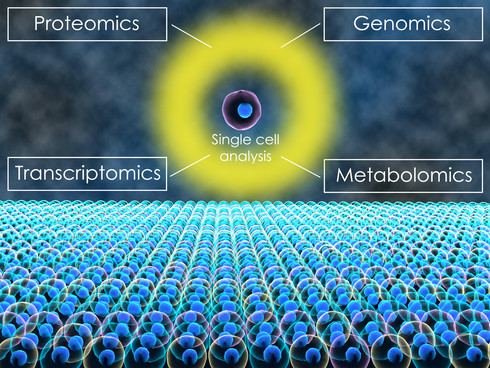
 Figure 1: A single blood draw contains multiple layers of biological information — genomic mutations, cfDNA fragmentation patterns, DNA methylation, cfRNA transcripts, and chromatin marks — each answering a different question about the tumor.
No single liquid biopsy signal captures everything a tumor releases into the bloodstream. Mutations tell you which genomic changes are present but miss cancers without druggable drivers. Fragmentomics detects genome-wide chromatin disruption but cannot resolve individual variants. Methylation reveals epigenetic programming. cfRNA captures active transcription. Chromatin marks from cfChIP-seq show which genes were active in the cells that released the DNA. The strongest evidence now comes from combining them. This guide walks through what each signal measures, what it adds, and how to design a multi-signal study that maximizes the biological information from every blood draw.
CD Genomics provides liquid biopsy solutions spanning cfDNA fragmentomics (low-pass WGS), targeted EM-seq methylation, HEBER-seq cfRNA sequencing, cfChIP-seq, and cancer panel sequencing.
TL;DR
- One signal is rarely enough. Mutations miss low-TMB cancers; fragmentomics cannot resolve variants; methylation alone loses tissue-context information. Multi-signal approaches consistently outperform any single modality in published 2024–2025 studies.
- The five signal types: Genomics (mutations, CNVs), fragmentomics (size, end motifs, nucleosome footprints), methylation (5mC patterns), cfRNA (transcript abundance, fusions), and chromatin marks (cfChIP-seq for histone modifications).
- Practical integration: Low-pass WGS provides both fragmentomics and CNV data from one library; adding methylation or cfChIP-seq on a second aliquot layers epigenetic information; cfRNA adds transcriptional context for tissue-of-origin and fusion detection.
- The 2025 evidence: SPOT-MAS Plus achieved 78.5% sensitivity at 97.7% specificity across 5 cancers by combining methylation + fragmentomics + 700 hotspot mutations. GUIDE/GutSeer reached AUC 0.921 with methylation + fragmentomics for GI cancers.
- What to do next: Start from your research question, not the technology. Define which biological layers answer it, then select the signal combination that covers those layers at the lowest complexity.
The Five Signals: What Each Layer Reveals
Each liquid biopsy signal originates from a different aspect of tumor biology. Understanding the biological basis of each signal — rather than treating them as interchangeable detection methods — is essential for choosing the right combination for a specific research question.
Signal 1: Genomics — Mutations and Copy Number Alterations
What it measures: Somatic single-nucleotide variants, small insertions and deletions, and copy number alterations in tumor-derived cfDNA. Detected via targeted panels at >1,000× depth or via low-pass WGS for genome-wide CNV profiling.
What it answers: "Does this sample carry a specific actionable mutation?" and "Are there genome-wide copy number changes?" Genomics is the most mature liquid biopsy signal, with well-validated bioinformatics pipelines and established variant calling benchmarks. Cancer panel sequencing at CD Genomics covers clinically and biologically relevant gene sets at the depth needed for reliable plasma cfDNA variant detection.
Limitations: Requires a mutation to be present in the panel's gene set. Low-TMB cancers, early-stage tumors with few detectable mutations, and cancers of unknown primary may produce false negatives from mutation-only approaches. Mutations alone cannot determine tissue of origin.
Signal 2: Fragmentomics — Chromatin-Level Cancer Signatures
What it measures: Fragment size distributions, end motif frequencies (4-mer profiles), and nucleosome positioning patterns from low-pass WGS (0.1–6×). These features reflect the chromatin organization and nuclease activity in the cells of origin (Cristiano et al., 2019).
What it answers: "Is cancer present?" (independent of mutations), "Which tissue did it come from?", and "Is the tumor signal changing during treatment?" The CD Genomics cfDNA fragmentomics service provides genome-wide fragmentation profiles, end motif analysis, nucleosome footprinting, and integrated DELFI-style scoring.
Limitations: Cannot resolve individual sequence variants. Nucleosome footprinting requires 5–6× coverage for reliable quantification. Fragmentomics features are influenced by pre-analytical variables (collection tube, processing time, freeze-thaw cycles) and require rigorous QC. For a detailed comparison with targeted panels, see our guide on cfDNA fragmentomics vs targeted cancer panels.
Signal 3: Methylation — Epigenetic Programming of Tumor Cells
What it measures: 5-methylcytosine (5mC) patterns at CpG sites across the genome or at targeted regions. Methods range from genome-wide (WGBS, EM-seq) to targeted (targeted bisulfite sequencing, cfMeDIP-seq). Methylation patterns are tissue-specific and cancer-specific, making them particularly informative for tissue-of-origin and early detection.
What it answers: "Is the methylation pattern consistent with a specific cancer type?" and "Which tissue released this cfDNA?" Methylation-based approaches underpin several multi-cancer early detection tests. The GUIDE/GutSeer study used 1,656 targeted methylation markers combined with fragmentomics to achieve 81.5% sensitivity at 94.4% specificity across five GI cancers in an independent test cohort (Huang et al., 2025). CD Genomics offers targeted EM-seq for methylation analysis with low DNA input requirements compatible with plasma cfDNA.
Limitations: Bisulfite conversion or enzymatic conversion can degrade cfDNA, reducing library complexity and introducing fragmentation artifacts that may complicate combined fragmentomics analysis (Lin et al., 2025). Enzymatic methods (EM-seq) cause less damage than bisulfite and are preferred for combined methylation-plus-fragmentomics workflows.
Signal 4: cfRNA — The Transcriptional Layer
What it measures: Cell-free RNA in plasma, including mRNA fragments, long non-coding RNAs, microRNAs, and other small RNAs. cfRNA is released through both passive (cell death) and active (extracellular vesicle secretion) mechanisms, providing complementary information to cfDNA. HEBER-seq (HEK293-based Endogenous RNA sequencing) and other cfRNA workflows capture the transcriptional activity of tumor cells from plasma.
What it answers: "Which genes are actively expressed in the tumor?" and "Are there gene fusions or splice variants detectable in plasma?" cfRNA is particularly valuable for detecting gene fusions — where intronic breakpoints are spliced out of RNA but retained in DNA — and for monitoring expression of tissue-specific or drug-resistance transcripts. CD Genomics provides HEBER-seq cfRNA sequencing for transcript-level liquid biopsy analysis.
Limitations: cfRNA is less stable than cfDNA and requires strict sample handling (rapid plasma separation, RNase-free processing). Approximately 95% of plasma cfRNA is non-coding, ribosomal, or mitochondrial; extracellular vesicle enrichment increases the mRNA fraction. cfRNA quantity is typically lower than cfDNA and may require higher plasma input volumes.
Signal 5: Chromatin Marks — Gene Activity Imprinted on cfDNA
What it measures: Histone post-translational modifications (H3K36me3, H3K4me3, H3K27ac) on nucleosomes circulating in plasma, detected by cfChIP-seq (cell-free chromatin immunoprecipitation sequencing). Active genes carry H3K36me3 in gene bodies and H3K4me3 at promoters; these marks survive circulation and can be immunoprecipitated from plasma.
What it answers: "Which genes were transcriptionally active in the cells that released this cfDNA?" cfChIP-seq provides a personalized gene-activity profile from plasma without needing a tissue biopsy. Wang et al. demonstrated that combining cfChIP-seq with methylation and fragmentomics achieved 90.4% sensitivity for early-stage lung cancer at 83.1% specificity (AUC 0.94) (Wang et al., 2025). The CD Genomics cfChIP-seq service enables histone mark profiling from plasma samples.
Limitations: cfChIP-seq requires substantially more plasma input than fragmentomics or methylation alone (typically 1–4 mL depending on the mark). The technique is newer than other liquid biopsy modalities, with fewer published validation studies. Antibody quality and specificity are critical variables. cfChIP-seq data is most informative when combined with at least one other signal type.
 Figure 2: Comparison of the five liquid biopsy signal types — genomics, fragmentomics, methylation, cfRNA, and chromatin marks — showing the biological source, key readouts, and primary research applications for each.
Signal Combinations That Work: Evidence From 2024–2025 Studies
Individual signals have known strengths and weaknesses. The question is which combinations produce the largest gains for specific research goals. The 2024–2025 literature provides clear direction.
Fragmentomics + Methylation: The Core Combination
This is the most extensively validated multi-signal combination. Fragmentomics provides genome-wide chromatin-level detection independent of mutations; methylation adds tissue-specific epigenetic information. The GUIDE/GutSeer study directly compared the combined model against fragmentomics alone and found the integrated approach significantly outperformed (AUC 0.963 vs 0.887, p<0.001) (Huang et al., 2025). The practical advantage is that low-pass WGS and targeted methylation sequencing can be performed on aliquots of the same cfDNA library, minimizing additional sample requirements.
Fragmentomics + Methylation + Mutations: Maximum Sensitivity
Adding hotspot mutation analysis to the fragmentomics-plus-methylation core further improves detection. SPOT-MAS Plus combined methylation, fragmentomics, and 700 hotspot mutations in a single workflow and found that mutations contributed most in liver cancer (96.5% sensitivity for hotspot mutations alone) while methylation-plus-fragmentomics excelled where mutations were scarce (breast cancer 51.6%, gastric cancer 62.9% for mutations alone) (Nguyen et al., 2025). The three-signal combination reached 78.5% overall sensitivity at 97.7% specificity. The lesson: mutations fill the gap where methylation and fragmentomics are weaker, and vice versa.
Fragmentomics + cfChIP-seq: Chromatin-Informed Detection
The MERGE approach (Multi-Epigenetic Regulated Genes) integrated cfChIP-seq (H3K4me3), reduced-representation bisulfite sequencing, and low-pass WGS on the same cfDNA samples. By identifying genes where chromatin marks, methylation, and fragmentomics features all pointed toward cancer, the ensemble model reached 90.4% sensitivity for stage I lung cancer — substantially higher than fragmentomics-based models alone (Wang et al., 2025). cfChIP-seq adds cost and plasma volume requirements, but the gain in early-stage sensitivity is notable.
Fragmentomics + cfRNA: Complementary Detection Mechanisms
cfRNA captures actively transcribed genes — including fusion transcripts and expression-level changes — through both passive release and active secretion. This makes cfRNA complementary to cfDNA-based signals: cfDNA reflects primarily cell death, while cfRNA also reflects active cellular secretion. For studies where gene fusions or expression-based subtyping are primary endpoints, adding cfRNA to a cfDNA-based workflow (fragmentomics or panel) adds transcriptional context that DNA alone cannot provide.
Designing a Multi-Signal Study: Practical Considerations
Adding signals adds biological information — but also adds sample requirements, cost, and analytical complexity. The following framework helps determine which signals justify their incremental burden for a given research goal.
Sample requirements by signal combination
| Signal Combination | Typical Plasma per Sample | Library Preparations | Sequencing Runs |
|---|---|---|---|
| Fragmentomics only (low-pass WGS, 1–6×) | 1–2 mL | 1 (WGS library) | 1 (low-pass) |
| Fragmentomics + targeted methylation | 2–4 mL | 2 (WGS + EM-seq) | 2 |
| Fragmentomics + methylation + panel | 3–6 mL | 2–3 | 2–3 |
| Fragmentomics + cfChIP-seq | 3–6 mL | 2 (WGS + ChIP) | 2 |
| Full multi-signal (all five) | 6–10+ mL | 4–5 | 4–5 |
These are directional estimates. Actual requirements depend on the specific protocols, desired depth, and the expected cfDNA yield from the study population. Plasma volume is often the binding constraint — if total available plasma is limited to 2–4 mL per sample, prioritize the signals that directly answer the primary research question and consider banking an extra tube for future analysis.
When one signal is enough
Not every study needs multi-signal integration. A single signal is sufficient when: (1) the research question is narrow — for example, tracking a known EGFR T790M mutation during treatment, where a targeted panel is the right tool; (2) the cohort is enriched for a tumor type where a single signal performs well — liver cancer has high detection rates from hotspot mutations alone; or (3) budget and sample constraints preclude adding signals. In these cases, choose the signal that most directly answers the question and invest the budget in sample numbers and replicate robustness rather than signal breadth. For guidance on study design specifics, see our cfDNA fragmentomics study design guide.
When integration justifies the cost
Multi-signal approaches add the most value when: (1) the cohort includes multiple cancer types with variable tumor mutational burden; (2) early-stage detection is the goal, where any single signal may fall below the detection threshold; (3) tissue-of-origin identification matters for the study endpoint; (4) the study aims to discover biomarkers that combine genetic, epigenetic, and transcriptional features; or (5) maximizing negative predictive value is critical. In these scenarios, the incremental cost of a second or third signal is typically justified by the gain in detection sensitivity and biological resolution demonstrated in the 2024–2025 literature.
Decision Framework: Which Signals for Which Goal?
Use this framework to align your research question with the appropriate signal combination before contacting a provider.
| Research Goal | Recommended Signal Combination | Why |
|---|---|---|
| Track a known mutation during treatment | Targeted panel alone | Deep, quantitative variant-level data at known positions; fragmentomics adds marginal value for this narrow question |
| Multi-cancer early detection (broad screen) | Fragmentomics + methylation | Best-validated combination; genome-wide detection + tissue-specific methylation; GUIDE/GutSeer provides the template |
| Multi-cancer detection with maximum sensitivity | Fragmentomics + methylation + hotspot mutations | Three signals cover each other's blind spots; SPOT-MAS Plus model; mutations add power for high-TMB cancers |
| Tissue-of-origin for unknown primary | Fragmentomics + methylation (or cfChIP-seq) | Both fragmentomics (nucleosome footprints) and methylation provide tissue-specific patterns |
| Early-stage detection (stage I/II emphasis) | Fragmentomics + methylation + cfChIP-seq | MERGE model reached 90.4% sensitivity for stage I lung; chromatin marks guide fragmentomic feature selection |
| Gene fusion and expression-based subtyping | Fragmentomics + cfRNA | cfRNA captures fusions and expression; fragmentomics provides genome-wide detection backbone |
| Biomarker discovery (broad feature space) | Maximum affordable signals | Larger feature space increases the probability of identifying novel composite biomarkers; subsequent validation can down-select to essential signals |
| Treatment response monitoring (no known mutation) | Fragmentomics (DELFI-TF style) | Mutation-independent tumor fraction tracking from fragmentomes; proven in van 't Erve et al. 2024; add methylation if tissue-of-origin shifts are relevant |
 Figure 3: A decision flowchart for selecting liquid biopsy signal combinations — starting from the research question, branching through cohort characteristics, sample constraints, and study endpoints.
FAQ
Can I combine all five signals from a single blood draw?
In principle, yes — but plasma volume is the binding constraint. A single 10 mL blood tube yields approximately 3–5 mL of plasma. Fragmentomics (1–2 mL) plus one additional signal (1–2 mL) is feasible from one tube. Three or more signals typically requires multiple tubes drawn at the same venipuncture. The practical limit for most studies is two to three signals per timepoint. If multi-signal analysis is a priority, plan the blood collection volume and tube count accordingly during study design.
Which signal combination has the strongest published evidence?
Fragmentomics plus methylation is the most extensively validated combination in 2024–2025, with the GUIDE/GutSeer and SPOT-MAS Plus studies providing prospective and retrospective validation across multiple cancer types. The addition of hotspot mutations to this core combination (SPOT-MAS Plus model) produced modest but consistent improvements in overall sensitivity. cfChIP-seq and cfRNA are newer modalities with fewer published validation studies but are active areas of development.
Does enzymatic methylation sequencing (EM-seq) preserve fragmentomics features?
Enzymatic conversion (EM-seq) causes less DNA damage than bisulfite conversion and better preserves fragment length distributions, though some alteration of fragmentomics features still occurs. Lin et al. (2025) comprehensively evaluated the impact of WGBS on fragmentomic characteristics and documented systematic shifts in fragment size and end motif profiles after bisulfite treatment. If combined methylation-plus-fragmentomics analysis from the same aliquot is desired, EM-seq is preferred over bisulfite-based methods. Alternatively, split the cfDNA library — one aliquot for low-pass WGS (fragmentomics), another for methylation — to avoid conversion-induced fragmentation artifacts altogether.
How do I prioritize signals when budget or sample volume is limited?
Start with the signal that directly answers your primary research question. If the question is "does this sample contain cancer?", fragmentomics provides the broadest detection signal per unit of cost and plasma. If the question is "what specific mutation is present?", a targeted panel is the right primary tool. Add a second signal only when the first signal has a documented weakness for your cohort — for example, add methylation if tissue-of-origin matters, or add mutations if your cohort includes tumor types where fragmentomics alone shows lower sensitivity. The CD Genomics Liquid Biopsy Solutions team can provide feasibility guidance for specific signal combinations based on sample type and study goals.
What is the minimum plasma volume needed for a two-signal study?
For fragmentomics plus methylation from separate aliquots, 2–4 mL of plasma is typically sufficient (1–2 mL per signal). For fragmentomics plus cfChIP-seq, 3–6 mL is more realistic due to the higher input requirements of cfChIP-seq. These are directional estimates — actual requirements depend on the expected cfDNA yield from the study population, which can be substantially higher in cancer patients than in healthy controls. Discuss specific input requirements with your provider during project scoping and consider running a pilot with representative samples to confirm feasibility.
References
- Mining nucleic acid "omics" to boost liquid biopsy in cancer. Tivey A, Lee RJ, Clipson A, et al. Cell Reports Medicine, 2024; 5:101736. DOI: 10.1016/j.xcrm.2024.101736
- Genome-wide cell-free DNA fragmentation in patients with cancer. Cristiano S, Leal A, Phallen J, et al. Nature, 2019; 570:385–389. DOI: 10.1038/s41586-019-1272-6
- Cancer treatment monitoring using cell-free DNA fragmentomes. van 't Erve I, Alipanahi B, Lumbard K, et al. Nat Commun, 2024; 15:8801. DOI: 10.1038/s41467-024-53017-7
- Combination of Hotspot Mutations With Methylation and Fragmentomic Profiles to Enhance Multi-Cancer Early Detection. Nguyen THH, et al. Cancer Medicine, 2025; 14(1):e70575. DOI: 10.1002/cam4.70575
- GUIDE: a prospective cohort study for blood-based early detection of gastrointestinal cancers using targeted DNA methylation and fragmentomics sequencing. Huang A, Guo DZ, Su ZX, et al. Mol Cancer, 2025; 24:163. DOI: 10.1186/s12943-025-02367-x
- Cell-free epigenomes enhanced fragmentomics-based model for early detection of lung cancer. Wang, Guo, Huang, Li, et al. Clin Transl Med, 2025; 15(2):e70225. DOI: 10.1002/ctm2.70225
- Comprehensive evaluation of the impact of whole-genome bisulfite sequencing on the fragmentomic characteristics of plasma cell-free DNA. Lin Y, et al. Clin Chim Acta, 2025; 566:120033. DOI: 10.1016/j.cca.2024.120033
- cfOmics: a cell-free multi-Omics database for diseases. Nucleic Acids Research, 2024; 52(D1):D1144–D1153. DOI: 10.1093/nar/gkad777
Research Use Only. This article is for educational and informational purposes and does not constitute medical or diagnostic advice.